When you find out that your doctor has ordered a CT scan due to the doctor finding a mass or a problem in your body can be frightening and intimidating. You may quickly start thinking about the worst-case scenario, but here is an important point to remember: not all masses are dangerous. Learning to distinguish the difference between cyst and tumor results on imaging will make you feel more knowledgeable and less fearful about what your scan results will show. The way cysts and tumors present on the CT scans is different, and the difference in the presentation will guide the radiologists in what they are dealing with. Even though certain masses need to be kept under tight surveillance or additional assessment, a great number of them prove to be harmless cysts that do not demand treatment whatsoever.
What is the distinction between a cyst and a tumor on a CT scan?
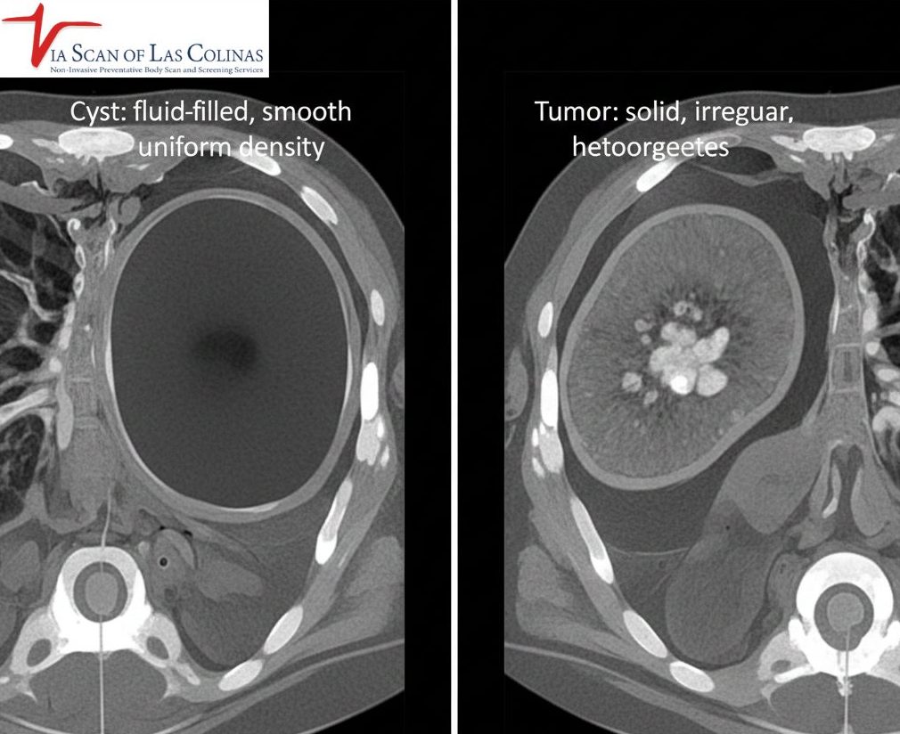
The very distinction between cyst and tumor on CT images is reduced to the contents of the mass. A cyst is a fluid-filled bubble – imagine it is a small water balloon in your body. In case of a CT, cysts are observed to be round and dark since they are filled with liquid, and not solid tissue. Tumours, on the other hand, are hard masses composed of real tissue or cells. Tumors do not appear the same on CT scans- they are usually brighter or more varied in appearance since solid tissue appears denser.
Can a CYST be Cancerous?
Most cysts are harmless and fully benign; however, some complex cysts with an abnormal appearance may need further examination to exclude the presence of cancerous alterations, and that is why professional interpretation of images is needed.
What would a cyst normally appear like on a CT Simple?
Simple cysts appear very characteristic on a CT scan and enable radiologists to identify them fast and be sure about them. Cysts are smooth, oval or rounded, and well delimited between the edges of the cysts; they are clean and neat looking and not irregular and jagged. Since they contain fluid, Cysts usually appear uniform in density on CT imaging, similar to fluid. Another characteristic that distinguishes them is that they do not enhance when contrast material is injected during scanning. Simple cysts have thin walls, including ultrasound-thin walls, so that they are barely visible, and the contents are homogenous, free of solid elements. All these typical characteristics inform radiologists that they are dealing with a benign and non-spreadable cyst that does not often need treatment or intervention.
Features that Indicates a Harmless Discovery.
- Round form: Circular or oval edges that are not irregular.
- Equal density of fluid: Dense or dark in all parts of the mass.
- Thin walls: Essentially, it is barely visible or very thin outer lining.
- None: Fails to brighten on administration of contrast dye.
- No hard parts: There are no solid parts or pieces of any tissue present.
- Borders: The cyst has clear edges, distinct borderlines between the cyst and the surrounding tissue.
- Uniform internal appearance: Even internal structure throughout.
What are the ways of identifying and assessing tumors on a CT scan?
Tumors appear very dissimilar to simple cysts in CT scans and the dissimilarity helps the radiologists assess the type of mass they are viewing. The solid tumors are brighter or more heterogeneous when compared to the dark and uniform cysts filled with fluid. In the issue of cyst vs tumor identification, radiologists seek masses that are irregular in shape, lack smooth edges or finger-like projections protruding into the surrounding tissues- things that cysts do not possess. Lots of tumours improve tremendously when contrast material is injected that is, they become bright on the scan as the solid tissue will absorb the contrast. Some mixed masses are not entirely solid or liquid and need close consideration. The ViaScan CT scan professionals in Irving, TX, offer the detailed scan that is required in the characterization and assessment of masses.
Solid vs. Mixed Mass Features
- Solid tumors: These are completely tissue based masses and appear brighter and more complicated on CT scanning with irregular contours, unequal density and enhancement of contrast- this also raises further medical examination to establish whether the tumor is benign or malignant.
- Mixed or complex masses: The masses are composed of solid tissue and fluid components, and they show partially dark and partially bright on CT scans, which are complex and thus it becomes more difficult to immediately identify whether the mass is benign and additional imaging or biopsy is needed to make a definitive diagnosis.
Does a CT scan effectively determine the benignity and malignancy of a mass?
CT scan provide exceptionally useful data on the masses but they cannot always be conclusive in diagnosing whether a tumor is cancer or not. In comparing cyst vs tumor results, CT imaging proves to be very successful in identifying simple and benign cysts that have clear fluid nature. In solid tumors, CT images reveal size, shape, location and relation to surrounding structures- all significant pointers to diagnosis. Some of the features involve increased risk: irregularity of borders, infiltration into surrounding tissues, quick growth between the scans, or unusual enhancement patterns. Yet, the question remains, is whether a cyst could be cancerous or whether it is a solid tumo and in this case, imaging alone may not always suffice.
Limits of CT Imaging
- Unable to tell the type of cell: CT is able to show the structure of the mass, but is unable to tell whether the cells are cancerous or not.
- Similarity in appearance: There are benign and malignant tumors which have similar appearances on imaging.
- Imaging findings should be correlated with the findings and medical history: Imaging findings must be correlated with symptoms and medical history.
- Biopsy is frequently required: Tissue sampling is the definitive diagnosis in the case of inconclusive imaging.
- Not amenable to microscopic disease: Since minuscule cancerous alterations are small, they can be missed in the CT scan.
- Variability of enhancement: Enhancement can vary both in benign and malignant masses.
- The follow-up can be required: Tracking scans over time can be used to assess growth patterns and behavior.
Choose Our Preventive CT Scan
Early Detection Saves Lives!
-
- Accurate
- Quick Result
- Affordable

Conclusion
When your doctor has found out that there is a mass or an abnormality, the first step in the right direction to know what you are talking about is to have proper imaging. Our state-of-the-art CT scan services at ViaScan in Irving, TX, provide good-quality images that healthcare providers require to make a good differentiation between cysts, tumors, and other results. Our professional interpretation and our well trained radiologic technologists guarantee the best imaging quality and will provide you with the clear answers that you need.