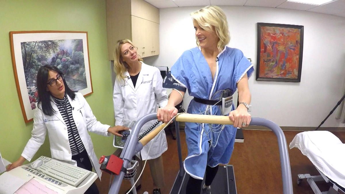
Nuclear Stress test utilize radioactive “tracers” such as Thallium and Cardiolyte to increase the clarity of a Nuclear Stress test. It’s purpose is to determine how well blood is flowing through the heart and is an indicator of the mechanical condition of the heart. The problem is a nuclear stress test will miss 100% of early plaque buildup and may only detect an issue once plaque has reached 70% blockage. The tests are graded normal, abnormal or equivocal (unknown). Basically they are a guess. Nuclear Stress test can not see plaque in arteries, so it only detects issues once damage has occurred to the heart.
With nuclear stress testing, false positive rate is 20% meaning that 20% of the people are sent to the hospital for cardiac catherization costing upwards of $20,000 and subjecting the patients to a 5% chance of death only to find out there are not major blockages. Conversely, nuclear stress test show a 15% false negative rate meaning that 15% of the people are sent home and told there are not issues only to discover they have major blockage that results in death or permanent heart damage via heart attack.
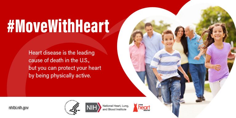
Prevention is predicated on the belief that treating potentially life-threatening conditions at the earliest possible point allows you to maintain good health rather than trying to regain it once it’s lost. At ViaScan heart plaque is seen as small as 1 mm in size, providing years or decades advanced warning of a buildup of plaque that may result in a heart attack or require surgery