
MRI vs CT Scan for Brain Imaging
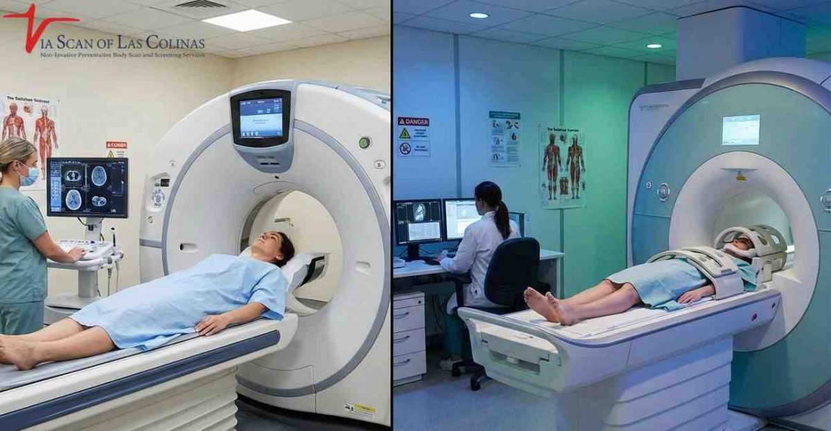

When a doctor recommends brain imaging, you may wonder which test is better: an MRI or a CT scan. Neither is universally better than the other. Each imaging method has different strengths, and the right choice depends on your symptoms, medical history, and what your healthcare provider needs to examine. Understanding the differences between these two tests can help you know why one may be recommended over the other.
Which provides better brain imaging, MRI or CT scan?
Neither MRI nor CT is universally better for brain imaging. MRI provides greater soft-tissue contrast and is often preferred when doctors need detailed images of brain tissue, such as when investigating tumors, multiple sclerosis, or certain neurological conditions. A CT scan is faster and is commonly used in emergencies to check for bleeding, skull fractures, and other urgent problems.
For example, after a significant head injury, a CT scan is often the first imaging test because it can quickly identify serious bleeding or fractures. If a doctor needs more detailed information about brain tissue, an MRI may be recommended. The choice depends on the patient’s symptoms and the medical question being investigated.
Key Diagnostic Differences
- Speed: A brain CT scan usually takes only a few minutes, while an MRI examination often takes around 30–60 minutes. Actual appointment times vary.
- Soft-tissue detail: MRI provides better contrast between different types of soft tissue, making it useful for detailed brain imaging.
- Bone visualization: CT scans show skull bones and calcifications more clearly.
- Availability: CT scanners are generally more readily available in emergency departments.
- Cost: CT scans are often less expensive than MRI scans, but prices vary by facility and location.
- Patient comfort: CT scans are usually quicker and less confining. Open MRI scanners may help some patients who experience claustrophobia, although availability and image quality vary by scanner.
When Each Scan Is Preferred
- MRI is the choice for: brain tumors, multiple sclerosis, ischemic stroke detection (especially beyond the hyperacute phase), detailed brain structure evaluation, pituitary disorders.
- CT scan of choice: Head trauma, possible bleeding in the brain, skull fracture, initial emergency stroke assessment to rule out hemorrhage, screening of abnormalities.
- The benefits of CT include emergencies, metal implants in patients, and fast screening.
- MRI benefits: elaborate neurological assessment, soft tissue disease, sequential imaging of known diseases.
Which brain conditions can be optimally identified through MRI?
MRI is good at identifying conditions of the soft tissues of the brain and the internal organs. It is a type of imaging in which extremely detailed images of your brain are produced without radiation by using intense magnets and radio waves. MRI, especially with contrast, may show issues that may not be visible using the other tests of imaging. MRI is commonly used by neurologists when exploring symptoms such as chronic headache, vision alteration, memory loss, or unexplainable neurological symptoms, which need to be examined in detail through the use of brain tissue.
Soft Tissue and Nerve Detail
MRI has an unparalleled visualization of both the white and gray matter of the brain whereby the structures that cannot be seen well via CT scanners are seen clearly in MRI. This is a thorough imaging that assists the physician to determine inflammation, infection or minute tissue changes which may give a clue of disease. MRI is able to visualize the nerve routes of the brain and this is very effective when assessing disorders that influence the functionality of the brain. The technology also enables the doctors to view various types of tissues depending on the amount of water content and chemical structure and also displays abnormalities which may not be detected by other imaging systems.
Tumors and Neurological Disorders.
In cases where doctors suspect that the brain has a tumor, MRI is normally the imaging test of choice since it reveals the size, location and attachment of the tumor to the other structures of the brain in an astonishing manner. The MRI is also the gold standard in the diagnosis and monitoring of multiple sclerosis as it makes the characteristic lesions of the brain and spinal cord visible. In conditions such as Alzheimer disease, MRI assists physicians in determining how the brain volume and tissue deteriorates with the course of time. Also, MRI is known to give detailed images in the detection and follow-up of aneurysms, disorders of the pituitary gland and assessment of congenital brain conditions that may not be seen in CT images.
In which cases does a CT scan become superior in the evaluation of the brain?
A CT scan to the brain turns out to be the better option in cases where there is urgent outcome especially during an emergency. In cases where one has sudden severe symptoms such as loss of consciousness, acute confusion or head trauma, doctors require an answer within minutes rather than 30 to 60 minutes which an MRI would demand. They seek such answers fast, and CT scans are the provider of time-sensitive treatment decisions to the medical teams. The CT scans are indispensable in the emergency departments because of the speed advantage that could save lives literally.
What is the difference between the MRI and CT scans in terms of safety and radiation?
Among the major differences in MRI and CT scans is the exposure to radiations. MRI involves magnetic fields and radio waves; no radiation at all, it is therefore safe to be used again and again in instances when it is medically required. A CT scan on the brain also involves X-ray radiation but modern CT technology has greatly minimized the level of exposure to X-ray radiation than the older machines. A brain CT uses ionizing radiation, and the risk from a single medically necessary scan is generally low, which is why it is used when the diagnostic benefit outweighs the risk. [2] Your medical practitioner is very mindful of the radiation exposure, particularly for children or patients who need scans on a regular basis.
Radiation Considerations
- CT radiation exposure: A brain CT uses ionizing radiation. For most patients, the risk from one medically necessary scan is generally low, and doctors order it when the benefit of an accurate diagnosis outweighs that risk. [2]
- MRI safety profile: MRI does not use any radiation, so it may be preferred when repeated scans are needed over time. Its safety for people with metal implants or devices depends on the specific implant; many are MRI-compatible under certain conditions, so always tell the staff about any implant. [1]
Conclusion
The decision between making an MRI and CT scan of the brain will be determined by your medical circumstances, and the medical practitioner will prescribe the right test to undertake. If your doctor orders a brain CT, ViaScan of Las Colinas in Irving, TX, offers CT imaging, overseen by John Duncan, Ph.D., whose 20-plus years in the field have focused on helping thousands of people take charge of their health. Each scan is read by a radiologist, and the report goes to your doctor.
References:
[1] U.S. Food and Drug Administration — MRI (Magnetic Resonance Imaging): Benefits and Risks. https://www.fda.gov/radiation-emitting-products/mri-magnetic-resonance-imaging/benefits-and-risks
[2] U.S. Food and Drug Administration — What are the Radiation Risks from CT?. https://www.fda.gov/radiation-emitting-products/medical-x-ray-imaging/what-are-radiation-risks-ct