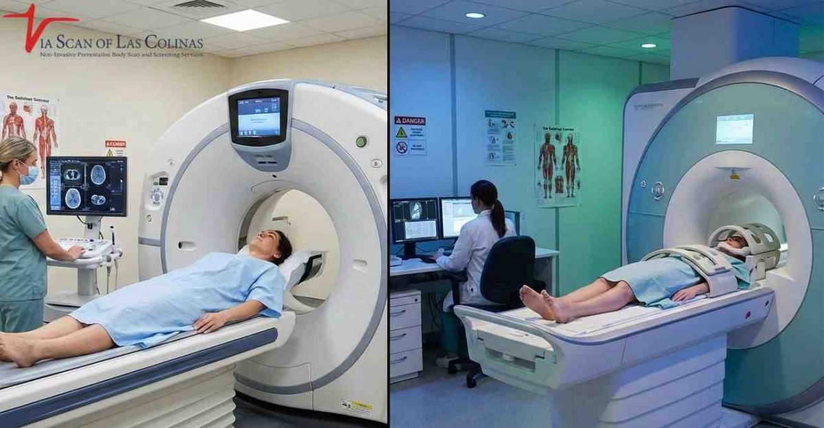

While an MRI machine can be used to examine different areas of the body, most MRI examinations focus on one specific region that a doctor wants to investigate. Imagine an MRI machine as a specialized camera. It does not capture images of the entire body at once unless a whole-body examination is specifically requested. Instead, it typically focuses on a particular area, such as the knee, brain, or spine. This is because many people undergo an MRI to investigate a specific concern, such as persistent shoulder pain or a headache that does not go away. However, the same technology can also be used to examine multiple areas of the body through a whole-body MRI protocol. Let’s make this as simple and easy to understand as possible.
Is there a difference between a whole body MRI and an MRI of a specific area?
An MRI typically examines a specific region of the body, such as an arm, abdomen, or brain, because the examination is often requested to investigate a particular symptom or medical concern. However, if someone wants a broader examination of multiple body regions, the same MRI technology can be used for a whole-body scan. The examination usually takes longer and follows a different imaging protocol than a targeted MRI.
What MRI scan covers
The duration of an MRI appointment depends on the area being examined and the type of imaging required. A targeted MRI generally focuses on one specific body region, while a more comprehensive whole-body MRI examination may take longer because it covers multiple areas. The exact duration depends on the scanning protocol and the imaging facility.
Which parts of the body can be scanned using an MRI?
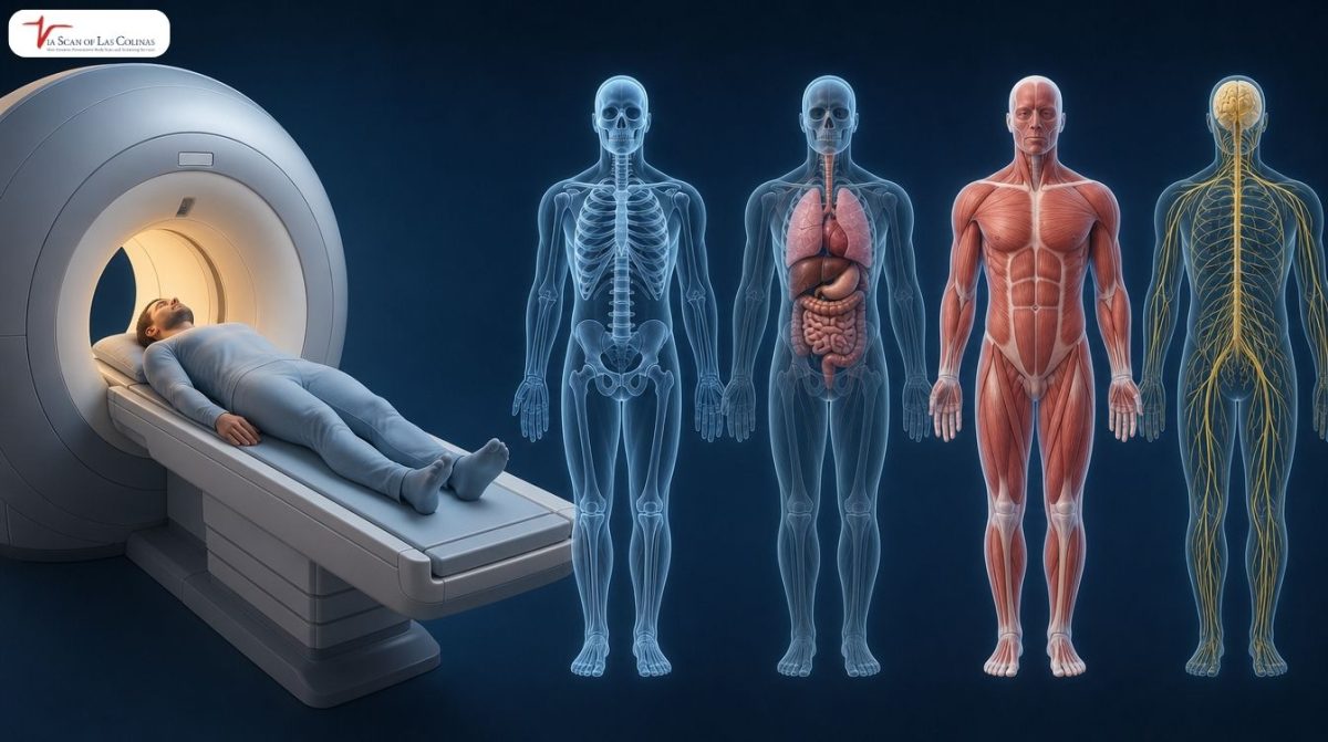
MRI technology can examine many parts of the body, including the brain, spine, heart, joints, muscles, and internal organs such as the liver. Because MRI uses magnetic fields and radio waves rather than X-rays, it is particularly useful for producing detailed images of soft tissues, including the brain, muscles, and internal organs [3].
Body Areas Examined by MRI
These are common areas inspected:
- Brain and spine
- Heart and chest
- On the belly and other organs such as the liver
- Knees, shoulders and joints
- Muscles and soft tissue
Can you have a Whole-Body MRI Scan to find out if you have health issues?
Yes, a whole-body MRI examination can provide images of multiple areas of the body during one examination rather than focusing on just one specific region. Some individuals choose this type of examination because they are interested in a broader overview of their health. However, it is important to understand that whole-body MRI screening is not a routine recommendation for every healthy individual. Its suitability depends on personal circumstances, medical history, and the purpose of the examination [2].
Whole-Body MRI Scans
If you’re considering a whole body MRI screening, here’s what to know:
- Visits numerous areas during one scan
- It could be used as a component of preventive test.
- Does not diagnose/treat anything on its own
- All results are checked by qualified staff
A whole-body MRI is not a magic solution and should not replace regular medical checkups or recommended health screenings. It may provide additional imaging information, but it also has limitations, including the possibility of incidental findings that require further investigation.
Whole-body CT scans, such as those offered at ViaScan of Las Colinas, are a different type of examination that uses X-rays rather than magnetic fields. The FDA notes that whole-body CT screening in people without symptoms has not been shown to provide more benefits than potential harms. Therefore, it is important to understand the differences between MRI and CT examinations and discuss their potential benefits and limitations with your doctor before proceeding [1].
What is the difference between a whole-body MRI and a targeted MRI scan?
A targeted MRI examines a specific body area, such as the knee or brain, to produce detailed images of that particular region. In contrast, a whole-body MRI uses a broader imaging protocol to examine multiple body regions during one examination. It provides a wider overview rather than concentrating exclusively on one specific area. The imaging coverage and level of detail depend on the protocol used and the purpose of the examination.
Whole-Body vs Targeted MRI
Imagine taking a picture of your entire backyard compared with taking a close-up picture of just one flower. A targeted MRI is like the close-up photograph: it focuses on a particular area that needs detailed examination. A whole-body MRI is more like the wider photograph, covering several areas during one examination.
Although whole-body MRI can examine multiple regions, it is not a substitute for dedicated imaging when a specific medical concern requires a more detailed examination. The American College of Radiology (ACR) states that there is insufficient evidence to recommend total-body MRI screening for individuals without symptoms, relevant risk factors, or a family history suggesting an underlying condition [2].
Neither approach is automatically better than the other. The appropriate examination depends on what the individual and their healthcare provider want to investigate, the person’s medical history, and the clinical reason for imaging.
MRI Services and Other Useful Scans
Imaging centers at ViaScan of Las Colinas also provide other imaging services, including heart scans, whole-body scans, and CT scans. A heart scan can provide information about the heart and surrounding structures, while a CT scan uses X-ray technology to produce detailed images of areas such as the chest or abdomen.
Whole-body imaging services may appeal to individuals who prefer to have multiple body regions examined during one appointment rather than scheduling separate visits for different areas. However, the suitability of these examinations depends on the individual’s circumstances and the type of information being sought.
These scans do not independently provide a complete medical diagnosis or treatment. Instead, they produce imaging information that can be discussed with a licensed healthcare professional who understands your medical history and can determine whether further evaluation is necessary.
Conclusion
Does an MRI scan the whole body? Most MRI examinations focus on a specific body area, although it is possible to examine multiple regions through a whole-body MRI protocol. MRI technology can provide detailed images of a particular joint, organ, or other body structure, as well as a broader view when a whole-body examination is performed.
Whether you are considering a targeted MRI or a whole-body examination, it is important to understand what the scan covers and what its limitations are. These examinations provide imaging information and should not be considered a replacement for regular medical checkups, recommended screenings, or professional medical evaluation.
Always consult a licensed healthcare professional to understand which imaging examination may be appropriate for your circumstances and what any findings mean for your health.
References
[1] U.S. Food and Drug Administration (FDA). Full-Body CT Scans: What You Need to Know. https://www.fda.gov/radiation-emitting-products/medical-x-ray-imaging/full-body-ct-scans-what-you-need-know
[2] American College of Radiology (ACR). ACR Statement on Screening Total Body MRI (April 17, 2023).https://www.acr.org/News-and-Publications/Media-Center/2023/ACR-Statement-on-Screening-Total-Body-MRI
[3] RadiologyInfo.org – American College of Radiology and Radiological Society of North America. Magnetic Resonance Imaging (MRI).https://www.radiologyinfo.org/en/info/mri
[4] Whole-Body MRI for Preventive Health Screening: A Systematic Review of the Literature. Journal of Magnetic Resonance Imaging.https://pmc.ncbi.nlm.nih.gov/articles/PMC6850647/