Kidney-Stone-Symptoms-and-Stages-1200x1200.jpg
06/Aug/2026

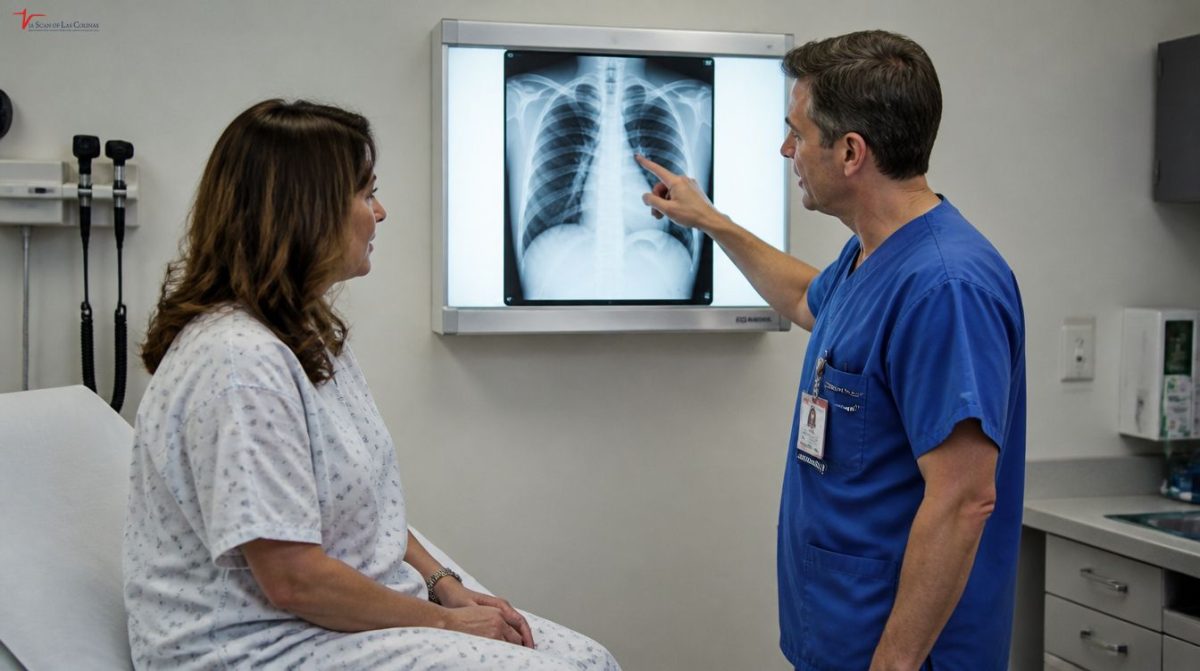
Symptoms of kidney stones vary with the progression of the stone’s formation, blockage, and passage down the urinary passages and will help to determine when to seek medical attention. Kidney stones are small rocks or stones formed from a buildup of minerals and salts in the kidneys. Some remain small and are not painful; some move and become painful. Knowing the signs of a kidney stone and the kidney stone stages can be beneficial when it comes to recognizing signs early on. Imaging is very important in monitoring the size and position of a stone in the urinary tract. 

What are the first symptoms and signs of a kidney stone?

Early signs and symptoms of a kidney stone are often overlooked, since a small stone might not initially cause pain. When the stone is growing or is starting to move, you may feel discomfort in your lower back or side. There may also be mild abdominal discomfort, cloudy urine or increased urination. There can be blood in the urine as well. A lot of people ask what causes kidney stones and some common reasons include dehydration, high mineral levels in urine, some specific diets, and family history. The information on kidney stones, and what causes them, will enable you to have a discussion with your health care provider about prevention. Further medical evaluation is recommended if symptoms persist or worsen.

Early warning signs to not overlook.

  • Some pain in the back or side
  • Frequent urination
  • Burning during urination
  • Cloudy or pink urine.
  • A slight stomach upset or nausea
  • A small amount of blood in the urine (also called hematuria)
  • An onset of symptoms that are getting worse gradually

What happens when a kidney stone passes down the urinary system?

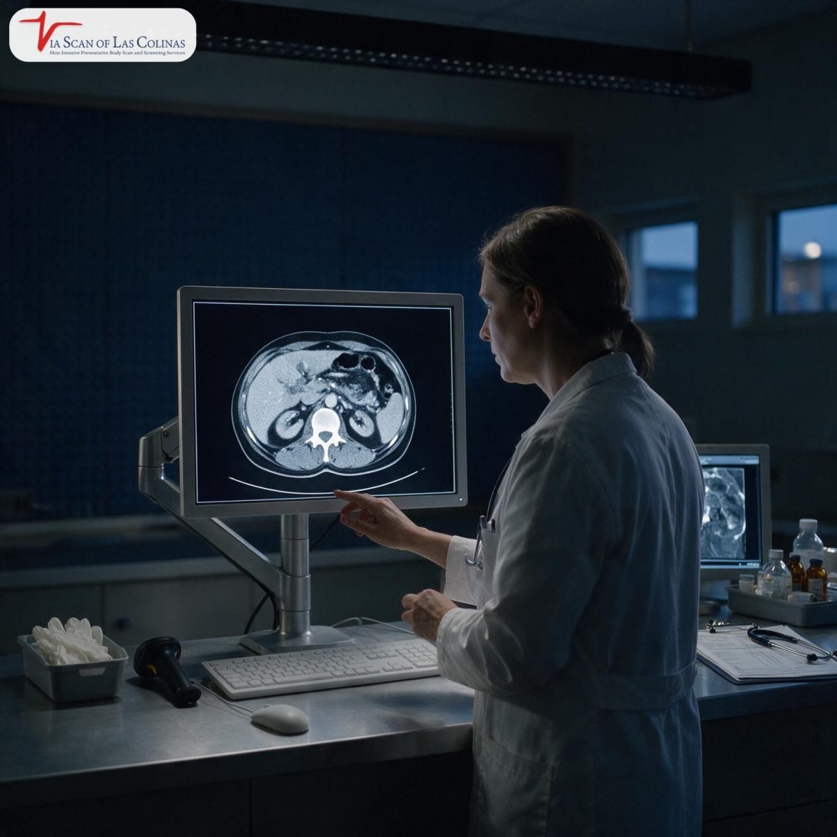
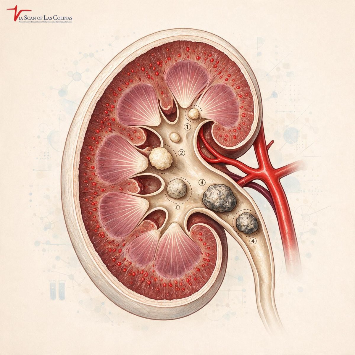
Kidney stones typically start in the kidney and pass through the narrow tube that transports urine from the kidney to the bladder (ureter). This trip tells about the various kidney stones stages. Sometimes it blocks the passage of the urine for a short while and then the pain is further increased. The pain may be intermittent as the muscles of the urinary tract keep pushing the stone. One of the most common questions is: How long does it take to pass a kidney stone? This will depend on the size, location, and the movement of your body. A kidney stone size chart can help a healthcare provider determine if a stone might pass or if it needs to be further evaluated. Over time, a CT Scan may be useful to track the stone’s progress. Advanced imaging services provided by ViaScan can help inform your health care provider’s discussions, and provide detailed images of the urinary tract.

Understanding the Different Stages of Stone Movement

  • A kidney stone develops in the kidney.
  • Over time, Stone grows bigger.
  • The stone is in the urinary tract.
  • The pain may become stronger and weaker.
  • Urines may be affected, becoming temporarily reduced in volume.
  • Stone moves into the bladder.The stone passes into the bladder.
  • During the process of urination, stone finally leaves a body.

What are the symptoms of kidney stones during each of the stages?

Often the pain shifts as the stone gets passed. The pain can start in the back or side and spread downwards towards the lower abdomen or the groin. The kidney stone passing symptoms vary and may indicate the stone is passing through the urinary tract. Some also experience more urination, burning, nausea or blood in the urine. How long it takes to pass a kidney stone depends from person to person, as each stone moves at a different rate. Doctors may use a kidney stone size chart along with imaging to gain a better understanding of how the stone is moving. A whole-body scan is used for preventive health screening, but a targeted CT Scan may be ordered if you have symptoms of a stone and your healthcare provider wants to learn more about the size and location of the stone. Medical evaluation should not be substituted with these imaging tools, which are useful for tracking changes.

Other common symptoms include: Pain Location, Urinary Changes

  • Pain starts in the back and radiates to the groin.
  • Frequent urination.
  • There could be burning while peeing.
  • Blood may be visible in urine.
  • Vomiting and/or nausea can occur.
  • Kidney stone passing symptoms are different for different people.

When is it time to see a doctor for a kidney stone?

Most small stones can pass on their own with supervision from the doctor, but some symptoms are serious and must be treated right away. If pain, fever, chills, vomiting, or trouble urinating does not go away, it is never OK. All of these symptoms may indicate infection or blockage and require immediate assessment. If you don’t know if your symptoms are serious, seek help right away. Imaging can aid determine that the stone has moved or grown in size. Advanced CT Scan imaging offered by ViaScan can help physicians monitor and monitor the position and motion of stones. Their imaging services are designed to offer detailed diagnosis information, not treatment. Your findings can then be reviewed by a licensed healthcare professional who can suggest what might be needed in your case.

Symptoms and complications that may occur during an emergency.

  • Extremely painful that doesn’t go away.
  • Fever or chills.
  • Continuous vomiting.
  • Unable to pass urine.
  • Blood in urine (hematuria).
  • Infection that causes pain.
  • Seek immediate medical attention.
  • Try to use the advice of licensed health care providers.

Choose Our Preventive CT Scan

Early Detection Saves Lives!

    • Accurate
    • Quick Result
    • Affordable

Conclusion

When kidney stones pass through the urinary tract they can develop a variety of symptoms. Understanding the symptoms of kidney stones, kidney stone stages, symptoms of passing a kidney stone, and how long it will take to pass a kidney stone will help you in the event of any change in symptoms, and you’re able to act quickly. A kidney stone size chart and imaging may also be used by healthcare providers to better understand a stone’s movement. Advanced services of CT scan and whole-body scan offered by ViaScan of Las Colinas could be helpful in giving patients and healthcare providers an understanding of what is going on over time and aid in medical decision-making.


Early-Detection-of-Neck-Lumps-1200x1200.jpg
27/Jul/2026

If a neck lump does not go away within the first 2 to 4 weeks, it is a good time to consider medical imaging to evaluate the lump and always sooner rather than later. If you’ve seen a lump on the side of your neck, felt something at the back or had trouble with swallowing and swelling, it’s important to know what’s going on. Neck lumps can be caused by a variety of things, some of which may be harmless, but others will require immediate attention imaging is one of the best ways to distinguish the differences quickly.

What are Neck Lumps and Common Causes of Lumps or Swelling in the Neck?

Neck lumps are any abnormal swelling or mass that is felt or seen in the neck area. Knowing the answer to the question of why does a lump appear in the neck begins with an understanding of the many structures within that small space, which can give rise to a lump for quite different reasons: lymph nodes, salivary glands, thyroid tissue, muscles and blood vessels.

Lymph Nodes, Cysts, Thyroid Nodules, and Other Causes

  • A swollen lymph node is the most common cause of a lump on the side of the neck and is usually a reaction to an infection, usually from a cold, strep throat, or dental abscess, that clears up when the infection is gone.
  • Thyroid nodules are growths in the thyroid tissue on the front of the neck—usually they are benign, but sometimes they need imaging and labwork to determine the significance or nature of the nodule.
  • A cyst, such as a sebaceous or branchial cleft cyst, is a non-cancerous fluid-filled sac that can form anywhere in the neck, at any age, and is usually soft, movable and grows gradually without causing much pain.
  • Neck swelling can also be caused by benign conditions such as lipomas and salivary gland enlargement, although sometimes the appearance of these masses can be mistaken for more serious conditions that need to be clinically assessed by a professional.

When should a neck lump be investigated with medical imaging?

The best question anyone with neck swelling can ask themselves is when to worry about a neck lump. Not all lumps need to be immediately studied by imaging — some traits make professional evaluation truly time sensitive.

Warning signs and factors that may indicate further testing are warranted.

  • A lump that has existed for more than 2-4 weeks and in which imaging and evaluation are warranted, especially if there has not been a recent illness, is a consistent clinical presentation that imaging and evaluation should be performed without delay.
  • A lump on the back of neck or side of neck that is hard, fixed, or is a growing lump is worrying rather than a soft, movable lump  imaging can characterize a lump far more precisely than physical exam.
  • Any neck lump causing difficulty in swallowing, hoarseness, unexplained weight loss or night sweats with swelling significantly increases the clinical concern and warrants immediate medical and imaging work up.
  • Pattern of neck lump cancer symptoms  in adults over 40, a persistent, firm enlargement without pain, especially if the person has a history of smoking or drinking — is always suspicious and should be carefully evaluated with imaging studies.

Which Imaging Tests Are Used To Evaluate Neck Lumps?

There are several ways to assess a neck lump and the most appropriate one will depend on what is suspected to be the cause of the lump, where the lump is located and how much additional information is required apart from what is found on physical examination.

How Ultrasound compares to CT Scans and MRI.

The first imaging test used for a lump on the side of the neck is usually ultrasound, which is quick, gives no radiation, and can be used to help determine the characteristics of lymph nodes and distinguish solid masses from fluid-filled cysts.

  • When cancer symptoms are suspected in a neck lump and the involvement is suspected to be deep, CT scans can give detailed cross-sectional imaging—very useful for this purpose.
  • MRI is excellent for soft tissues, does not use radiation and is preferred in the imaging of deeper neck compartments and salivary glands where high resolution differentiation of tissues is important.
  • PET-CT may be performed when malignancy is suspected, to find out if the tissue is growing and for better detection of any other areas of involvement than the main lump.

How Can Medical Imaging Help Detect Potentially Serious Neck Conditions Early?

Medical imaging can detect serious neck problems early because it shows abnormalities that cannot be seen on a physical exam, such as small, deep masses, early thyroid changes, and enlarged lymph nodes that don’t yet cause surface swelling. Imaging data adds to the clinical picture to make decisions about when to be concerned about a neck lump clearer. Early imaging is a valuable tool for persistent or concerning swelling, as there is often a definitive answer as to what is causing a lump in the neck.

If you prefer to see things in detail and from an anticipatory perspective, ViaScan of Las Colinas provides non-invasive CT scans and whole-body scans that scan multiple regions of the body at once. A whole-body scan can detect abnormal findings in the neck along with other internal abnormalities, aiding in early, informed discussions with your health care provider. If you have a neck lump and are having trouble swallowing or have a lump on the back of your neck – talk to a licensed professional before adding preventive imaging to your health plan.

Choose Our Wellness Body Scan

Early Detection Saves Lives!

    • Accurate
    • Quick Result
    • Affordable

Conclusion

Neck lumps occur regularly and are generally non-cancerous but if the lump is persistent or accompanied by symptoms, it is important to seek professional advice as soon as possible. ViaScan of Las Colinas provides professional CT scan and whole body scan services to people with a proactive approach towards health awareness, knowing early is better than knowing later. Seek professional assistance early; early assessment always results in better outcomes.


How-To-Treat-Swollen-Lymph-Nodes-1200x675.jpg
06/Jul/2026

Neck lymph nodes can get swollen pretty often; it’s usually part of what the body does when there’s a small infection, or even just a short-lived immune response. Still, figuring out when this kind of swelling is more benign and you can handle it at home, versus when it needs professional evaluation, is just as important. Swollen lymph nodes aren’t really a condition by themselves, more like a sign that your immune system is paying attention to, and reacting to, something going on. This blog gives you the answers to all these questions and more, along with some of the natural strategies that can help your recovery and when they may persist and some things that should always be taken seriously.

What causes neck lymph nodes to swell, and when are they harmless?

The most common cause of swollen lymph nodes in the neck is a response by the immune system to an infection in the area. Usually non- threatening, and will pass as soon as the cause is resolved. They are a concern if they continue, increase, or if they are accompanied by other, unexplained symptoms.

Some common infections and other potential triggers:

  • The most common reasons for swollen lymph nodes under jaw one side are common colds, flu, strep throat, and ear infections; when the immune system enlarges nearby nodes to fight off the invading bacteria or virus.
  • Swollen lymph nodes under jaw one side may indicate dental infections or an infected tooth, because the nodes next to the infection are most likely to be swollen first and most noticeable with the presence of the bacteria.
  • Swollen lymph nodes in neck one side may be due to an infection of the skin, insect bite or cut in the area near the neck and head, which is a localized reaction that usually goes away when the skin infection is treated completely.
  • In less frequent instances, swollen lymph nodes in the neck could occur due to autoimmune disorders, some medications or more serious underlying health issues such as early-stage neck cancer, so any unusual swelling should be checked.

What are the Natural Remedies for Swollen Lymph Nodes in Neck?

The swollen lymph nodes in the neck may be a result of a mild infection or a temporary immune response, and can be supported by natural home care. All these techniques have the effect of helping the body heal, but they will not directly treat the underlying cause.

Home Care Strategies for Recovery:

During the recovery phase, it is important to rest, drink plenty of fluids and use a warm compress to help ease the pain and boost the immune system. Anti-inflammatory foods, avoiding physical stress, and stress management can also set the stage for the body to heal mild lymph node swelling over time.

How long does it take for swollen lymph nodes to go back to normal?

If neck swelling is due to a minor infection, it will usually go down as the infection clears in 2-4 weeks. Nodes that persist in their enlargement beyond these limits, or which enlarge further during the course of the illness, should be investigated by a health care worker.

Rehabilitation Factors and Expected Healing Time

Sometimes, a mild swelling just shows up from a common infection, and it tends to settle on its own within 2-4 weeks. The whole recovery thing depends on how intense the underlying reason is, the overall immune defense, plus age, and also what kind of care was actually given. If the lymph nodes are swollen on only one side of the neck and they are still there for more than four weeks,without any obvious explanation , then you should seek medical advice right away.

What symptoms suggest that swollen lymph nodes may need medical evaluation?

Some signs of an inflamed neck lymph node could point to a more serious underlying issue (instead of just a routine infection) — and they really do warrant a professional assessment and managed approach, not just continued at-home care.

The signs that you should never overlook.

  • If these lymph nodes grow larger over several weeks after the infection has gone away, they need to be assessed urgently by a licensed health care professional.
  • Swollen lymph nodes, which are hard, fixed, or rubbery to the touch, under one side of the jaw, are one of the recognized warning signs and should be professionally evaluated and imaged.
  • If a person has unexplained weight loss, fatigue, night sweats, or a fever with neck lymphadenopathy, they really should be carefully assessed for a systemic illness, and a detailed physical examination needs to be done.  
  • If the lymph nodes on one side of the neck are swollen and they do not look infected, cold or generally “sick”, especially in adults over 40, then they should be carefully evaluated, and early stage swollen lymph nodes in neck cancer or other serious underlying problems should be ruled out as soon as possible.
  • Any node that is greater than one centimeter in diameter and has not been explained after more than four weeks should be treated professionally and may require appropriate imaging to accurately determine the cause of the node.

If you’re concerned about a larger area of your body that may be affected, especially if you have swollen lymph nodes that don’t go away ViaScan of Las Colinas provides non-invasive preventive imaging services such as a whole-body scan that covers the neck, chest, and abdominal area. These are awareness resources used for information and discussion with your health care provider, not as a substitute for emergency medical services. When swelling continues, seek professional help and consider taking proactive steps by getting imaging.

Choose Our Wellness Body Scan

Early Detection Saves Lives!

    • Accurate
    • Quick Result
    • Affordable

Conclusion

Tonsils that enlarge (swell) in the neck are typically harmless and will usually go away on their own, but they are definitely a sign to watch. Natural care helps with recovery for those with mild swelling, and if swelling is persistent, worsening, or accompanied by symptoms, seek prompt professional care. If in any doubt, do it early. At ViaScan of Las Colinas, people can get professional preventive imaging services without waiting for symptoms to become a concern, if they want to know what’s going on inside their bodies before it creates problems.


Lump-Under-Armpit-in-Women-and-Men-1200x655.jpg
25/May/2026

Finding a lump under your armpit can feel alarming, and yet most of the time it is explainable and usually not serious at all. So if you spotted a small lump sort of low in the armpit, or you noticed a larger one that appeared more suddenly, you will probably want to figure out what it might be, and then decide when it is smart to see your doctor. Below is a loose overview of the most typical causes, the warning signs, and how healthcare professionals can help you with different imaging options that are available for a lump under the armpit. 

What is the most common reason for a lump under the armpit in both women and men?

Lymph nodes, those little glands that help your immune system, live in the armpit area. These nodes swell in response to an infection, inflammation, and any changes within the body. A lump under the armpit in females, or a lump under the armpit in males, is often about the same kind of causes, and most of the time it is not malignant or anything cancer-like. 

infections, enlarged lymph nodes, and a few other health issues 

  • A smaller bump in the armpit is most frequently tied to swollen lymph nodes. That can happen when your immune system is busy dealing with something, like a cold, the flu, a skin infection, or even a seemingly “minor” cut on the arm. In these situations, the lump will often fade on its own once the infection clears, and the swelling goes down. 
  • Another fairly common idea is ingrown hairs or skin irritations, especially in places that get shaved. You may see a small red bump that seems to sit just near the skin’s surface. This can appear in both sexes, and it generally calms down with time if you stick to gentle skincare and try not to keep rubbing the area.
  • In the softer tissue of the armpit, it can happen that there are non-cancerous lumps, like cysts, or sometimes lipomas. A cyst is a sac filled with fluid, and a lipoma is a slow-growing mass of fat. Both are soft and movable and are usually not painful. 
  • The swelling of the lymph nodes around the armpit can be temporary and it comes from an allergic reaction or from the body’s response to a vaccination, sort of like a delayed kind of signal. This is pretty well documented after certain shots given in the upper arm, and it usually calms down within a few weeks.

When is a lump in the armpit more concerning? 

Most armpit lumps are generally harmless, but there are signs that sorta point you should not sit around and wait for things to “settle.” There are a few clues; if you recognize them can help you decide when it’s worth getting checked, not just hoping it fades. For example, if you notice a big bump under the armpit, or if it stays put for a long time without any movement, there are a few features to pay attention to.

Signs of trouble requiring medical care.

  • If the lump is firm, sessile (unmoving) and increasing in size then it should be seen by a doctor.
  • Benign lumps are soft and move slightly when touched. If the lump is hard and feels immovable and it seems to be growing larger over time, medical evaluation is recommended.
  • Any lumps that come with symptoms that are unexplained, like a loss in weight, night sweats, constant tiredness, or fever, should be discussed with a health care provider. 
  • When combined with a little lump under the armpit in females or males, these systemic symptoms can be a sign that the lymph nodes are reacting to something that needs an investigation.
  • If a lump does not go away in 2-4 weeks, it should not be ignored. Swelling of the lymph nodes due to infection will go away when the infection is cured. 
  • If a lump has not gone away in a month for a known reason, it is something that should be assessed by a professional.

Are Swollen Lymph Nodes Under the Armpit connected to cancer?

Yes, sometimes, swollen lymph nodes under the armpit are related to some types of cancer, such as breast cancer. Cancer cells that spread to the underarm lymph nodes can form a breast cancer lump under the armpit. Most lumps in the armpit area, however, are not associated with cancer. If any lump is detected in the imaging report, it should be examined and tested by a healthcare provider to determine the cause of the lump.

Benign Causes: Potentially Serious Causes
Swollen lymph nodes because of an infection Persistent lymph node enlargement, especially if there is no infection around Hard , immovable, quickly growing lump Lymphoma or other lymphatic conditions
Ingrown hair or folliculitis, that kind of irritated bump Lipoma, a fatty tissue growth, softish and slow A lump linked with unexplained weight loss or fever 
A cyst from a blocked gland often feels smooth or slow-moving Vaccine-related swelling can happen, but unusual persistence should be checked. 
An allergic reaction can also swell areas, but it typically comes with other symptoms and fades Metastatic cancer that affects the lymph nodes

How do health care providers determine the cause of armpit lumps?

When you visit your doctor about a lump under your armpit,  either a small lump under armpit that’s been there for weeks or a large lump under your armpit that you noticed recently your doctor will go through the evaluation process. The goal is to better understand the possible cause of the lump and determine whether additional testing is needed. Your health provider will answer all your concerns about why do I have a lump under my armpit. 

Physical Exam and Med Evaluation

  • Often, an initial physical examination is the first step. Your doctor will examine the lump to check whether it is movable, hard or tender, and will ask you about any recent infections or illnesses, vaccinations, and when the lump appeared. A physical examination helps physicians evaluate the characteristics of the lump and possible underlying causes.  
  • Imaging tests may be suggested if a lump is not adequately investigated by physical examination. A computed tomography (CT) scan can help doctors find out the size and appearance of the lymph nodes and nearby tissue. Las Colinas’ ViaScan provides non-invasive CT scan and whole-body scan services, which can complement a physician’s wider health evaluation process and offer further insights on imaging that could assist in determining next steps in a person’s healthcare.

If a physician suspects that any imaging or physical tests are useful to pursue, a biopsy or blood test may be ordered. The tests are conducted by specialist medical providers and are used to further detail the cause of persistent or problematic lumps.

Choose Our Wellness Body Scan

Early Detection Saves Lives!

    • Accurate
    • Quick Result
    • Affordable

Conclusion  

Whether you’re dealing with a lump under the armpit in a woman, or a lump under the armpit in a man, the biggest thing is not to just brush it off, ok. Most lumps end up being benign, a healthcare professional can properly evaluate the cause. is to get a proper medical evaluation. If that lump has hung around for more than a few weeks, it looks like it’s getting bigger, or it appears together with other symptoms, then it’s smart to talk with a licensed healthcare provider, without any delay. 

For people who really care about preventive health awareness and prefer more subtle and non-invasive screening choices, ViaScan of Las Colinas provides professional imaging services, like CT scans and whole-body scans, which are reviewed by experienced professionals. These imaging services are meant to encourage health awareness, not replace a clinician’s evaluation or any necessary medical treatment.


Symptoms-of-Low-Potassium-in-Females-1200x670.jpg
18/May/2026

Potassium is an essential mineral that helps support normal body functions; your muscles, your heart, your nerves. When they become too low, your body begins to send signals that you are quick to ignore as being tired or stressed. In particular, knowing how to identify the signs of low potassium before it develops into more severe health-related issues, and knowing when to take a test, may make a significant difference towards detecting a deficiency before it leads to additional and more serious health-related complications. And now shall we go through it all in a simple and clear manner?

What Are the Typical Symptoms of Low Potassium in Females?

Low potassium, also known as hypokalemia, is a condition that affects women of all ages and can be mild, moderate, or severe, depending on the extent to which the levels have decreased. Understanding how to know whether there is a low level of potassium begins with the most common early symptoms.

Early vs. Severe Warning Signs.

  • The initial symptoms include continuous fatigue, muscle weakness, mild cramping, constipation, and generally experiencing physical heaviness that is not relieved with rest or hydration.
  • Heart palpitations, a large amount of muscle twitching, numbness or tingling in the limbs, and pronounced weakness, which disrupts normal daily activities and function, are severe warning signs.
  • Ignoring early symptoms of low potassium can lead to abnormal heart rhythms and severe muscle abnormalities, making it crucially important to recognize the early symptoms of low potassium and establish a medical diagnosis of low potassium.

What Are the Causes of Low Levels of Potassium in Women?

Low potassium results when the body loses more potassium than it gains, or when underlying disorders interfere with how the body absorbs and retains this vital mineral. There are a number of reasons that are especially pertinent to the health of women in particular.

Diet, Medications, and Underlying Conditions.

  • One of the most prevalent and avoidable causes of low potassium in women is poor dietary intake, especially those who are low in fruits, vegetables, and whole foods. Crash dieting and restrictive eating patterns are associated with a high risk of deficiency in the long term.
  • Drugs such as diuretics, laxatives and some blood pressure medications cause the kidneys to secrete excess potassium, making women on these medications especially significant subjects of regular monitoring and routine blood tests.
  • Coexisting conditions like eating disorders, chronic diarrhea, vomiting, kidney disease, and hormonal imbalances, such as increased levels of aldosterone, directly interfere with the normal maintenance of adequate levels of potassium regardless of dietary intake.

What Are the Effects of Low Potassium on the Body and Health in General?

The direct role that potassium plays is in the mechanism by which the muscles contract, the nerves transmit signals, and the heart retains its rhythm. As potassium levels drop, all these systems are impacted, sometimes in ways that seem unrelated to a mineral deficiency until appropriate tests prove otherwise.

Effects on Muscles, Heart and Nerves.

  • Some of the first and most reliable physical symptoms are muscle twitching and cramping, which occur due to the need for potassium in normal muscle cell functioning and electrical signaling throughout the body.
  • Heart palpitations occur when low potassium levels interfere with the electrical impulses that regulate heartbeat rhythm — a symptom that always requires immediate medical attention and diagnosis through blood tests.
  • Low potassium impairs nerve functioning, causing tingling, numbness, and in severe cases, severe weakness that affects coordination and physical abilities beyond simple fatigue.
  • When potassium levels decrease, digestive activity slows down, which explains the bloating and constipation many women complain about but may not connect with potassium deficiency.

At What Stage Should a Woman Be Tested for Low Potassium Levels?

It is important to learn when to monitor potassium levels, as it is just as important as recognizing the symptoms. A basic blood test, known as a basic or comprehensive metabolic panel, measures potassium levels directly and helps provide a medical diagnosis on which clinical decisions are based. Do not hesitate to test before symptoms become serious.

Medical Symptoms That Require Testing.

  • Constant muscle weakness, cramping, or muscle twitching with no apparent cause that does not resolve with rest or hydration over several days.
  • Heart palpitations or any feeling of irregular, rapid, or fluttering heartbeat that is new, recurrent or accompanied by dizziness or shortness of breath.
  • Unexplained fatigue and digestive problems, such as constipation that lasts longer than a few days without an obvious dietary trigger.

Women who regularly take diuretics, blood pressure medications, or laxatives should ask their doctor about regular potassium monitoring since these medications can influence potassium levels before symptoms become visible.

Choose Our Full Body Scan

Early Detection Saves Lives!

    • Accurate
    • Quick Result
    • Affordable

Image

Conclusion

Having blood tests to monitor mineral levels in your body is an important part of overall health awareness. Understanding what is happening inside your body at a deeper level can help support preventive care. ViaScan of Las Colinas provides preventive imaging services such as whole-body scans and CT scans to complement overall health monitoring plans. These services do not replace clinical diagnosis or physician-directed care but can support proactive health awareness as part of a broader preventive strategy. Always discuss proper testing and monitoring with your healthcare professional based on your unique health needs.


Why-Does-My-Stomach-Hurt-in-the-Morning-1200x670.jpg
11/May/2026

This happens more often than most people understand because it creates confusion when someone experiences a stomachache during a time they should not be eating. Multiple factors cause morning stomach cramps because people fail to maintain their regular daily activities and their bodies undergo different digestive processes. The first step needs to start with learning how your body functions during sleep. The blog presents common reasons for stomach pain that occurs in the morning and describes when patients should rest and when they should seek medical help.

What are the reasons that stomach pain happens more often during morning time?

Your body remains active during your sleep. Your body continues to operate because your digestive system works continuously and your hormones fluctuate and your stomach acid moves through your body. Internal processes start functioning when the first light of dawn arrives between three and four o’clock. This explains why people experience stomach cramps during the early morning hours which happen before their first meal. If someone experiences slow digestion throughout the night it leads to incomplete digestion of their previous evening meal which results in morning bloating and pressure. Your body starts to process the nighttime activities as soon as the day begins.

Overnight changes in the digestive system and hormones.

  • Your body’s natural waking-up cycle turns up the production of cortisol in the morning. Some of these hormones can stimulate stomach acid levels, leading to stomach irritation, cramping or discomfort, particularly in those with sensitive digestive systems.
  • Overnight digestion process is slower, hence the food is digested more slowly during the sleep. This slower tendency may lead to excessive gas, bloating or leaving undigested food in the stomach for longer than usual and may not be comfortable in the morning.
  • People who experience morning stomach cramps more than three times each week should see a doctor to receive an official medical assessment. 

The digestive problems include acid reflux as well as gas and indigestion.

  • Acid reflux is when stomach acid comes up through the esophagus. This can be aggravated by lying flat all night and may be the first symptom of the day, such as burning or stomach pain.
  • Bloating and gas form at night when bacterial action occurs in your gut while the bacteria are going to work on undigested food. This trapped gas can lead to cramping, pressure and overall discomfort, which is most evident in the morning when you first get up and start to move around.
  • Indigestion (dyspepsia) may persist from food consumed the day before. It can be a diffuse sense of ache, pressure or indigestion in the upper abdomen which is more noticeable on an empty stomach in the morning.

In some cases, upper abdominal discomfort may also be felt near the rib area. Learn more about common causes of pain under the ribs above the stomach and when it may require medical attention.

Does the morning stomach pain happen because of Stress or Anxiety?

Yes, absolutely. The gut and brain has a direct link between the gut and the brain. Your digestive system is affected by your mind when your mind is stressed, even when it’s just a little bit stressed like when you’re really busy. People with anxiety experience their most intense stomach problems during the morning hours, which occur before they eat breakfast. This is not “imagined suffering. The situation proves to be a genuine and actual physical response. The American Psychological Association recognizes gastrointestinal distress as a symptom which links anxiety and chronic stress with their relationship to anxiety disorders.

What eating habits or foods can cause stomach pain?

The foods you consume together with your eating patterns create stomach pain which occurs after you wake up. Most people practice this behavior without understanding its existence. People who eat dinner after midnight experience stomach pain because this practice has become their normal routine. People who eat large meals during the evening hours force their digestive systems to operate beyond normal limits because their bodies should be resting. Your body will produce painful reactions because it cannot process the duration of your eating breaks. You can achieve positive results through simple dietary changes that you can make to your evening meals.

  • Late night stomach pain is commonly associated with eating 2-3 hours before going to bed. It is important for your tummy to empty sufficiently after eating, otherwise you might experience discomfort, bloating and acid reflux the next morning which is a result of lying down too quickly after a meal.
  • High fat, spicy foods and processed foods are harder to chew and digest and tend to be more irritating to the stomach’s lining. These foods can lead to acid, and may slow down food passing, making a morning upset more possible, particularly for those with sensitive digestion.

It’s also possible to experience stomach pain in the morning if you go without dinner completely. When the stomach is empty, it makes acid without anything to break down, causing stomach cramps in the morning, nausea or a burning sensation after getting up.

Choose Our Full Body Scan

Early Detection Saves Lives!

    • Accurate
    • Quick Result
    • Affordable

Image

Conclusion

Periodic morning stomach upset is often not a cause for concern. Sometimes an imaging test is ordered in conjunction with a more comprehensive examination. An abdominal CT scan can help doctors detect changes in the structure of the digestive organs that are not seen in a routine examination. 

As part of a health awareness approach, ViaScan of Las Colinas provides non-invasive preventive imaging services such as whole-body scans and CT scans. These scans may assist a physician’s evaluation and provide information about health but are not a substitute for the physician’s diagnosis or treatment. When stomach pain keeps occurring in the morning, it’s always best to talk to your health care provider first.


Cancer-Pictures-of-Swollen-Lymph-Nodes-in-Armpit-1200x670.jpg
04/May/2026

Swollen armpit lymph nodes can be due to a lot of things, including a small infection and something that actually needs to be examined by a doctor. The distinction between a lump to monitor and one to investigate promptly is precisely what this blog is there to assist with. We are going to pace what to seek, what auxiliary symptoms are important, and when to cease waiting and seek professional assistance.

What are the Swollen Armpit Lymph Nodes that are a result of cancer?

The first step to any educated health choice is to know what to seek. Although the diagnosis of the meaning of a swollen lymph node is always left to a doctor, there are both visual and physical attributes that certified medical experts always raise concerns as to the need to seek further investigation, especially when it comes to the causes of swollen lymph nodes in the armpit that can be severe.

Appearance and Indications of danger.

  • Hard, firm, or rubbery feel, which does not move when palpated easily unlike soft, mobile swollen lymph nodes that are more typical of infection than cancer lymph node swelling differences.
  • Enlargement of the area beneath the skin resulting in a noticeable raised area or lump in the armpit not before.
  • The skin changes around the lump with redness, dimples or abnormal texture – an event that is best checked with the doctor immediately.
  • Asymmetry – swelling in one armpit and no visible associated infection or injury on the same side of the body.

What Do You Think to Do Tell When a Swollen Lymph Node in the Armpit May Be Serious?

This is among the most significant questions that any person with the query when to worry about armpit lump must get answered. It is the physical appearance of the swelling, both in relation to the duration of the swelling and the presence of other symptoms that doctors consider when determining whether a lymph node should be investigated further.

Benign and Concerning Swelling: Differences.

  • The difference between infection versus cancer lymph node swelling is usually limited to the texture and movement, as infection related lymph nodes are usually soft, tender and movable whereas worrying nodes are usually hard, fixed and pain free.
  • The node that is related to breast cancer metastasis to lymph nodes armpit tend to be hard and non-tender and do not disappear in a few weeks, as the swelling caused by infection usually does.

What Are the Common Symptoms of Swollen Lymph Nodes that are caused by cancer?

The presence of a swollen lymph node in itself is half the story. Serious causes of lymph node swellings in armpit are usually followed by other symptoms that, when combined, would give a better clinical impression that your physician would consider.

Pain, Changes in Size, and Systemic Signs.

  • One of the systemic manifestations that predisposes when to worry about armpit lump to the list of urgent questions to be analyzed is unexplained weight loss and constant swelling of the armpits.
  • In conditions not necessarily falling into the category of infection vs cancer lymph node swelling, night sweats, chronic fatigue, and a fever with no apparent source of infection are common with lymph node involvement.
  • Enlarged lymph node when it progressively increases in size over weeks instead of decreasing is one of the physical signs that causes of swollen lymph nodes in armpit may need imaging and clinical evaluation.

What is the length of time a swollen armpit lymph node should last before going to the doctor?

Time is among the most practical advice on when to be concerned with armpit lump situations. Swollen lymph nodes usually heal in two to four weeks when the infection is removed. Persisting, growing, or emerging nodes without an apparent reason are of another scheme, and should be evaluated by a specialist.

How Long and At What Time It Concerns.

  • A physician should consider any enlarged armpit lymph node which has not diminished in size after two to four weeks – especially when there is no infection or trauma to account for the enlargement.
  • Lymph node spread Breast cancer that has spread to the armpits can manifest itself as painless, persistent swelling since lumps that last longer than one month without any apparent cause should always be subjected to professional imaging evaluation.
  • Swelling of an armpit lymph node during days to weeks (with or without pain) is an emergency indication to consult a doctor without delaying to the next routine check-up.

Choose Our Wellness Body Scan

Early Detection Saves Lives!

    • Accurate
    • Quick Result
    • Affordable

Conclusion

One of the most active health choices is to know what your body is like on the inside before symptoms get too serious. ViaScan of Las Colinas provides high-end preventive imaging services such as whole-body scans, CT scans, which are created to facilitate early health awareness as an element of a wider preventive plan.

The services are not a replacement of clinical diagnosis or physician-directed care – however, they can offer useful internal information that can be used to conduct more informed discussions between patients and their medical professionals.


Advanced-Kidney-Cancer-Diagnostic-Imaging-_-ViaScan-1200x800.jpg
16/Mar/2026

Kidney cancer is relatively common among adult cancers and is usually silent and does not produce any visible indicators of its presence at early stages, and it is difficult to detect without effective medical examination and radiography. Learning the process involved in diagnosing and identifying kidney cancer makes you appreciate the need to get regular health checkups particularly in cases where you are at risk of developing such risk factors as smoking, obesity, high blood pressure, or even family history. Timely diagnosis of kidney cancer is a significant aspect in the treatment process and that is why the use of sophisticated imaging is such an important factor in ensuring that kidney abnormalities are detected in good time to prevent them. 

What are the methods through which physicians detect kidney cancer at early and advanced stages?

Diagnosis of kidney cancer entails a review of the medical history, physical examination, laboratory tests, as well as advanced imaging studies, which present kidney abnormalities. Numerous cancers of the kidney are incidentally identified at the time of imaging to show some other cause as an early tumor is not likely to have symptoms. Whenever the kidney cancer symptoms are observed, i.e., blood in the urine, chronic back or side pain, unexplained weight loss, or fatigue, the symptoms may indicate a more advanced disease, which needs urgent examination and prolonged diagnostic tests to see the stage of the cancer and the available treatment options.

Primary Medical Examination and Signs of Kidney Cancer.

At the time of the initial examination, physicians enquire about symptoms of kidney cancer such as blood in urine (hematuria), chronic pain in the back or lower abdominal side below the ribs, unexplained weight loss, fever, fatigue, or palpation, an abdominal mass. A swelling or an abdominal mass may be detected on physical examination but this is not common in early stage cancer of the kidney. The majority of kidney cancers are silent and are detected accidentally in abdominal imaging due to other conditions at an early phase, and therefore, screening of health condition and diagnostic imaging is important to detect the risk factors.

Diagnostic Tests used for Confirmation

After the suspicion of kidney cancer, physicians test to diagnose and identify the extent of the cancer. Several imaging procedures such as CT scan of kidney cancer, MRI, and kidney ultrasound test give detailed images of the kidney structures showing the size, location and characteristics of tumors. Blood tests and urine tests evaluate kidney functioning and detect abnormalities that may be the indication of cancer. In certain cases, tissue biopsy can be done so as to study the cells under a microscope, but imaging can in most instances of kidney cancer suffice to make diagnosis and implement treatment.

What are the commonly used imaging tests to identify kidney cancer?

The modern imaging is the new kidney cancer diagnosis method where one can view a detailed kidney structure that depicts the presence of masses, tumors, and other abnormalities that are not directly seen or detected by the physical examination and blood test only. Kidney cancer CT scan is the most widely and valid imaging technique but MRI and kidney ultrasound test offer useful diagnostic information in certain circumstances. The professional CT scan services offered by ViaScan provide high quality imaging that are required to detect renal cancer and fully assess the kidney.

CT Scans, MRI, and Ultrasound

  • CT scan of kidney cancer: CT scans give excellent visual representations of cross-sectional pictures of kidney tumors with good level of clarity – the CT scan is enhanced with contrast that shows the blood supply to the tumor and differentiates between cancerous and benign cysts.
  • MRI imaging: Magnetic resonance imaging has better soft tissue contrast that can be used in cases where CT findings are inconclusive or in patients who cannot be administered CT contrast dye especially when determining the spread of tumors into blood vessels.
  • Kidney ultrasound test: This is an image test that involves the use of sound waves to provide real time images that help differentiate fluid filled cysts from solid masses – this is usually the first test to be performed on the kidneys when the abnormalities are suspected but not as detailed as CT or MRI.

The way Imaging Discovers Tumors in the Kidneys.

  • Mass identification: Imaging clearly reveals abnormal growth or malformation in kidney tissue in that it distinguishes between solid tumors and fluid-filled benign cysts that do not necessitate treatment.
  • Size and location mapping: The detailed images indicate the exact size of the tumor, its location in the kidney, and its connection with the surrounding organs – this is very important when treating the tumor.
  • Staging information: Imaging defines where kidney cancer has spread outside of the kidney onto the lymph nodes, blood vessels, or other organs nearby to the kidney – necessary to identify the stage of the cancer, and the way to treat it. Imaging findings can also help physicians understand how fast kidney cancer spreads and whether nearby organs or lymph nodes are already involved.

How long does it take to die from kidney cancer

The progression of kidney cancer varies greatly depending on the stage at diagnosis, tumor aggressiveness, and overall patient’s health condition. Early stage kidney cancers that are detected incidentally often have a much slower progression, and timely treatment can significantly prolong survival. Developed kidney cancer, particularly stage 4, can spread quickly to distant organs such as the lungs or bones, which may shorten life expectancy if untreated.

It is more important to note that each patient’s course is unique and survival depends on factors such as age, kidney function, response to treatment, and the presence of other health conditions. Regular monitoring and early intervention are key to managing the disease and improving outcomes.

What is the use of blood and urine tests in the diagnosis of kidney cancer?

Although the imaging can give a visual confirmation of kidney masses, blood and urine tests can be used to give deepening diagnostic information on the functioning of kidneys and health condition overall. These laboratory tests do not actually diagnose kidney cancer but assist in the diagnosis of abnormalities that need further investigation and give baseline health data that will be critical in the planning of treatment. Complete diagnostic assessment of kidney cancer requires evaluation by use of imaging as well as laboratory tests which are best done at an early stage.

Diagnosis of Abnormal Function of the Kidney.

  • Blood urea nitrogen (BUN) and creatinine: An increase of these values reflects kidney dysfunction which could be as a result of cancer of the kidney tissue.
  • Complete blood count (CBC): Discloses anemia, which is a frequent symptom of kidney cancer, or the presence of high red blood cell counts (polycythemia) that is occasionally caused by kidney cancer.
  • Liver function tests: The abnormal results can give an indication that kidney cancer has metastasized to the liver, although this is not the main task of these tests.

Evidence for Continued Testing.

  • Urinalysis: Reveals blood in urine (hematuria) – a frequent symptom of kidney cancer that would then lead to imaging to establish the source of bleeding.
  • Blood calcium level: Increased calcium (hypercalcemia) occasionally points to high grade kidney cancer that causes hormones that influence calcium metabolism.
  • Lactate dehydrogenase (LDH): High levels could be an indication of aggressive kidney cancer, but this is a nonspecific marker that needs to be confirmed by imaging.

Choose Our Preventive CT Scan

Early Detection Saves Lives!

    • Accurate
    • Quick Result
    • Affordable

Conclusion

Early diagnosis of kidney cancer enhances success rates and survival with a high degree of success. We offer specialized CT scan services and whole-body scan options that are at par with those offered in ViaScan, Irving, TX, to assist in diagnostic services of renal cancer in infancy and renal health checks using diagnostic imaging services.


What-is-the-most-Common-reason-for-a-CT-scan.jpg
16/Jun/2025

You have heard your doctor say that you require a CT scan, and suddenly, your mind is jamming. Just what is this scratch test? What is the purpose of it? And most importantly, what is the most frequently occurring cause that would make the doctor order such a type of scan?

CT scans may be something that one can easily think of as terrifying; however, when creating this impression, one is referring to one of the most useful tools available to doctors today. Imagine them to be something like the super-powered cameras that can look inside your body, and they do not even need to cut you open. We’re going to dive in and look at everything you want to know about these amazing medical images!

What is a CT scan? Why Is It so Common?

Computed Tomography, or just CT. Imagine it as a special X-ray, but with multiple pictures taken from various angles. Consider, for example, that you would like to find out what is inside a gift that has been wrapped in paper but has not been opened yet. An X-ray is akin to shining a flashlight at a specific angle – you can still see some shadows, but the entire image will not be visible. A CT scan is the equivalent of dozens of flashlights being shone in all directions, allowing a person to see inside and out of everything.

Why Are CT Scans So Popular?

They’re Fast

The average procedure on most CT scans requires only 10-30 minutes. It’s not as much time as you’d need to watch your favourite TV show!

They’re Detailed

CT scans can detect what is not visible even in regular X-rays. They are able to see the problems hiding behind the bones, or in other places which are not easily visible.

They do not hurt

CT scans do not cause any harm, unlike some medical tests. All you need to do is lie on a table and let the machine do its job.

They Assist Doctors to Make Rush Decisions

A CT scan can give doctors a quick insight into what is wrong with the patient, especially when such a patient visits the emergency room with critical symptoms.

What Is the Most Common Reason to Get a CT Scan?

To examine problems in the abdomen is reason number one which convinces doctors to order CT scan products. This involves exploring organs such as your liver, kidneys, pancreas and intestines. There is a 30% chance that CT scans will be used to examine these vital body parts.

What is the reason behind the prevalence of belly scanning? Your abdomen would be like a busy neighbourhood where many important organs are packed together. When your belly hurts, you cannot be too sure which part of the vicinity is causing it without giving it a closer scrutiny.

Why Does a Doctor Order a CT Scan?

 Although belly scans are most popular, there are many other reasons, as your doctor wants to have a little gadget inside your body:

Brain Scans and Head Scans

These are overly common, as well! The brain CT scans are ordered by a doctor when individuals experience the following:

  • Unrelenting bad headache
  • Light-headedness or dizziness, Dizziness or balance problems
  • Memory issues
  • Accident or fall, head injury

Chest Scans

The place that houses your heart and lungs is your chest. Physicians may request a chest CT scan as a measure to check:

  • Breathing problems
  • Chest pain
  • Non-productive coughing
  • Lung infections

The screening for cancer

CT scans are scavengers in the hunt for cancer cells. They use the ability to assist doctors:

  • Identify the tumours that may be hiding
  • Check whether the therapy against cancer is effective
  • Ensure whether it has spread to other parts of the body due to cancer.

Taking a glance at Injuries

CT scans can be used to quickly demonstrate when you are involved in an accident or if you hurt yourself:

  • Broken bones
  • Internal bleeding
  • Organs destruction
  • Blood vessel issues

Wellness and Prevention

The number of people undergoing CT scans, even when they do not feel unwell, is increasing. These wellness scans can identify problems early enough to prevent them from becoming severe.

What Do I Need to Do to Prepare for My CT Scan?

Preparation of a “CT scan” is generally quite simple! This is what you must know:

Check Your Rules of Eating

In some CT scans, you will need to avoid eating for several hours prior to the scan. Your physician will inform you about the necessary steps, such as fasting before a blood test – not an exciting prospect, but not difficult, either.

Take plenty of water.

Drink plenty of water, unless your doctor advises otherwise. This helps the kidneys function more efficiently and, in some cases, makes the scan clearer.

The Day of Your Scans

Do not Wear Tight Clothes

Opt for clothes that lack metallic zips, buttons, and undergarments. Metal appears in CT pictures and may prevent the view. Plump into pyjamas or sweatpants!

Take out Metals

Remove jewellery, watches, and all other pieces of metal. Even minor items such as bobby pins will influence the pictures.

Carry Your Medical Data

Carry a list of your medications and scan results from the past. It would be comparable to a person showing up to school with their homework, a positive way of helping others complete their work more efficiently.

Contrast Dye

In some cases, doctors may want to apply a special type of dye to enhance the appearance of a specific portion of your body in the scan. And don’t worry – it is normal.

Drinking Contrast

Before you have a scan, you may be required to take a chalky drink. It doesn’t have a particularly good taste, but neither is it particularly bad. Just imagine it’s a strange kind of milkshake that helps your doctor see better.

IV Contrast

In other cases, they will inject the dye into your arm using a small needle. You may experience a warm feeling or a strange taste in your mouth for several minutes. There is nothing wrong with this!

Is the CT Safe? What Risks are involved?

This is likely the question that causes the greatest concern for most people, and it is entirely normal to feel anxious about it. What is the situation? “CT scans” are quite safe. Each year, millions of people acquire them without difficulties. There are virtually always more pros than minor cons associated with learning about the medical problems ailing your body.

Radiation

When you are flying, you will be exposed to a small amount of space radiation. When you undergo a CT scan, you receive approximately the same amount of radiation as travelling across the country a couple of times by plane.

Your Body can deal with it.

Natural sources of radiation, such as the sun and other rocks, expose your body to a small amount of radiation each day. CT scans only do a few more in the short run.

The Risk Is Very Minor

The risk of acquiring cancer as a result of a single CT scan is too minimal, almost equal to your risk of being mesmerised by lightning.

What do the Results Mean?

You should not find it puzzling to interpret your CT scan findings nearly 100% of the time. This could be what could befall it:

Normal Results

When analysis reveals that everything appears to be normal, your doctor will inform you that the scan will not reveal anything. That is good news!

Abnormal Results

In the case that something abnormal is detected in the scan, do not freak out! Not everything labelled as abnormal is inherently bad. It simply may imply:

  • It is just a small infection that can be cured easily
  • It is something which must be monitored over time
  • You will require another test to have a better picture

Follow-Up Tests

Your doctor may occasionally need to conduct additional tests to gather more information. This resembles taking a second opinion – it is used to ensure that they get the full picture.

Choose Our Preventive Heart Scan

Early Detection Saves Lives!

    • Accurate
    • Quick Result
    • Affordable

Conclusion 

Whether you are considering a wellness CT scan or your physician has prescribed it, ViaScan is ready to make the process as easy and informative as possible. We understand that undergoing any medical test can be intimidating, but it doesn’t have to be. Whether you are undergoing a scan at your doctor’s request, we will guide you through all the procedures. We are eager to address all your inquiries and clarify everything in simple, easy-to-understand language.


Difference-between-CT-scan-and-MRI-1.jpg
11/Jun/2025

You are in the doctor’s facility, and they say that you require a scan. Suddenly, your mind begins to race with questions. CT scan? MRI? What is the distinction? Will that be painful? Should I fast?  Don’t worry! We are going to make you an expert in scanning without giving you a headache. Just imagine this is your introduction to these wonderful machines that look inside your body like X-ray vision superheroes.

Why are there so many Scans, and what is so Popular about CT and MRI?

Your body is a complicated puzzle with numerous pieces. There are cases where doctors require additional equipment to visualise other parts.

You can imagine it like when you take photos. There are times when you use your phone camera to take impromptu shots. Then there are times when you require a complex camera with specific lenses to capture close-up images. The CT scans and the MRI machines are like those various cameras – each of them is truly excellent at demonstrating things inside your body.

The most popular are CT scans and MRIs, as they are the Swiss Army knives of medical imaging. They are able to visualise many different things and assist the doctors to know what is going on inside you without necessarily having to operate on you first.

What is the CT Scan vs. MRI Difference?

The plain facts are these: CT scans involve X-rays (the same kind of radiation used in medical imaging, such as having your picture taken, only with invisible light), whereas MRI scans involve magnets and radio waves (being in a large magnetic field that can see through the body).

CT Scan:

  • Involves X-rays which revolve around you.
  • Composite photographs in slices, as if they were slicing bread
  • Super quick – normally takes 5-10 minutes
  • You are put on a table which moves inside a large doughnut-shaped machine

MRI:

  • The category utilises strong magnets and radio frequencies.
  • Slices pictures as well, but in greater detail
  • Time-consuming – typically 15-45 minutes
  • Produces thunderous knocking and buzzing 
  • You are lying in a tube-shaped machine.
  • They are both fantastic at their work, simply in different ways.

Which is safer, a CT Scan or an MRI?

This is what worries you the most. The truth of the matter is that both are quite safe when used responsibly.

The CT scans involve a minimal amount of radiation, equivalent to flying across the country a few times. Your body can cope with this just fine; however, doctors take care not to subject you to too many CT scans within a short period.

MRI scans do not require any radiation. Radio waves and magnets are generally safe for most individuals. The only downside is that you cannot have any metal in your body (such as pacemakers or metallic implants) since the magnet is extremely powerful.

Your doctor will never order a scan that you do not need, and they will always ensure that the benefits outweigh the risks by far.

What is Best Diagnosed with Which Type?

This is the place where these machines can really demonstrate their superpowers!

CT scans are excellent when:

  • Fractures and injuries
  • Trouble in your chest, belly and pelvis
  • Finding infections
  • Fast examination of your organs
  • Emergencies where answers are required quickly by doctors

MRI scans excel at:

  • Your brain and your spine
  • Examining muscles, tendons and ligaments
  • The visualisation of soft tissues at close-up view
  • Locating issues in the knees and shoulder joints
  • Obtaining truly clear images when physicians must view small details

Most of us have questions like, Can a CT scan detect cancer? And the answer is yes. CT scans are excellent in detecting a wide variety of cancers throughout the body. They are able to detect tumours, whether cancer has metastasised, and assist the doctors in designing treatment. MRI is also excellent at detecting certain cancers, particularly those of the brain, spine, and soft tissues.

What About Contrast Dye – Do the Two Tests Use It?

Now and then, yes; now and then, no! Contrast dye is like having special paint in your body that helps some areas show up better in the pictures.

If you are undergoing a CT scan, you may be given a chalky fluid to drink or receive an injection into a vein. There’s nothing to be ashamed of – according to most people, it only makes them feel warm for a few seconds.

In the case of MRI scans, contrast is normally administered via an IV. It is of a different kind compared to CT contrast and tends to produce fewer reactions.

Not all do need contrast dye. Your physician will inform you whether you require it and the reasons. If you do, the technologists will answer all your questions and closely monitor you to ensure you are fine.

Are CTs or MRIs Ok in Children or Pregnant Women?

In children, both scans are safe when required. To help children remain still, medication may be necessary, particularly during an MRI, as it is a longer procedure. Most hospitals even have kid-friendly machines that have cool decorations and TVs to watch cartoons!

In case of pregnant women:

  • MRI has been seen to be safe in pregnancy, particularly beyond the first trimester
  • Due to the radiation, CT scans are normally not performed during pregnancy unless in cases of emergencies.
  • The doctors will always consider the benefits versus any risks and will opt for the safest route for both mother and baby.

The point is that doctors order such scans only when necessary, and they always choose the safest option for everyone.

Choose Our Wellness Body Scan

Early Detection Saves Lives!

    • Accurate
    • Quick Result
    • Affordable

Conclusion 

CT scans and MRI machines are two procedures that have superheroes with different powers in your medical team. One is quick and excellent in emergency cases, whereas the other requires time to provide detailed information. They are all safe, painless, and designed to help you maintain your health.

And remember, having a scan does not mean that there is something wrong; it only means that your doctor wants to conduct a thorough examination to ensure your health. It is as though you were given a detailed map of your body, and your medical team could treat you most effectively.

Want to know more about body health maintenance using state-of-the-art scanning? ViaScan offers wellness body scanning services to help you manage your health effectively. As the saying goes, with your well-being, knowledge is power!