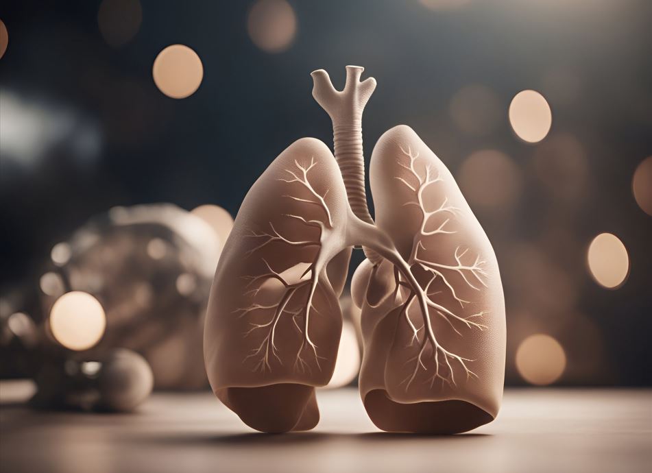
You’ve undoubtedly heard about gallstones if you haven’t personally encountered them. Who knows, maybe someone you know had their gallbladder removed? However, because gallbladder illness encompasses a variety of disorders that affect the gallbladder, the general issue might be perplexing. In this blog, we will dissect the functions, preventative measures, and primary ailments that are included in the gallbladder illness category. We’ll go over the symptoms and risk factors, as well as when scans are required. We will also discuss how different scanning facilities like ViaScan and their services, such as mobile CT Scans, have made life a little easier.
What is the role of the gallbladder?
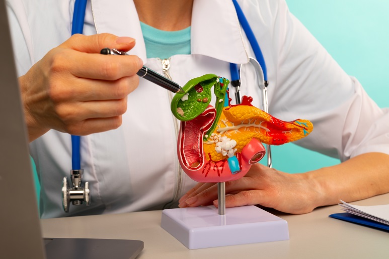
The gallbladder is a little, pear-shaped organ that sits just below your liver on the right side of your belly. It is essential for digestion. The primary gallbladder function is to hold bile, a substance your liver produces that aids in fat digestion. Your gallbladder discharges bile into your small intestine when you eat. More bile is needed for meals that include more fat. A gallbladder that isn’t functioning properly can cause many problems, and many disorders can impact it. The most frequent cause of malfunction is an obstruction that lowers bile flow. In more extreme situations, your gallbladder may need to be removed because it is not necessary for your body to operate.
What does your gallbladder do?
The gallbladder performs a number of vital tasks, including:
- To concentrate and store bile
- To react to hormones in the gut (such as cholecystokinin) in order to replenish and empty its bile reserves.
- To aid in controlling the bile’s composition, which includes its water content and bile salt content.
- To regulate the bile’s entry into the small intestine
- To constrict, bile is released into the duodenum, the first part of the small intestine, and the biliary system.
Which process can be primarily affected by gallbladder disease?
When the gallbladder isn’t functioning properly, bile accumulates in your blood rather than going to your small intestine to aid in digestion. You could experience digestive issues, particularly when it comes to breaking down fatty meals, as the breakdown of lipids in the small intestine depends on bile. Bile that accumulates in your blood will cause you to get unwell since it also transports poisons that your liver has removed from your body. Extreme pain might also be experienced from a diseased gallbladder.
Gallbladder function in the digestive system
The gallbladder purpose in the digestive tract is to produce Bile juice. Bile’s main job is to break down lipids. When the gallbladder contracts, bile is secreted. This takes happen in reaction to:
Gastric distension, or the stomach protruding because of a large amount of food in conjunction with fatty food content Cholecystokinin (CCK) is a hormone produced as food passes from the stomach into the small intestine. Bile begins to break down swallowed fat and fat-soluble vitamins as soon as it reaches the duodenum. It also helps to improve the ingested solubility of digested fat and facilitates its absorption.
What are the symptoms of a low-functioning gallbladder?
- Biliary colic, an infrequent soreness, is the most prevalent and mildest sign of proper gallbladder function and illness. An intense, persistent gripping or gnawing pain that often radiates to the upper back is felt by patients in the upper right abdomen, close to the rib cage. Acute cholecystitis, or inflammation of the gallbladder caused by stones or sludge blocking the duct, affects 1% to 3% of individuals with symptomatic gallstones. Similar to biliary colic, but more severe and long-lasting are the symptom Complaints of persistent diarrhea, nausea, and discomfort in the abdomen following meals are signs of chronic gallbladder disease.
Similar symptoms to those of gallbladder stones can be caused by stones stuck in the common bile duct. However, they can also cause: - Jaundice
- Lighter stools,
- dark urine
- Quick pulse and sudden
- decrease in blood pressure
- fever, chills, nausea, vomiting, and excruciating upper right abdominal discomfort
Where is the gallbladder located in the female body?
Studying human anatomy, it is very rare that a person’s organ may be located at a different location in the body. Male and female organs are both located in the same region. The gallbladder is a little, pear-shaped organ located just below your liver on the right side of your belly. A digestive fluid called bile is stored in the gallbladder and is expelled into the small intestine. Modern technological advancements also allow us to solve the mysteries behind the difference between male and female anatomy. Full body scan facilities such as Via Scan Of Las Colinas offer a number of scanning services, including mobile CT scans that promise clear images of the internal structure and functioning of human beings.
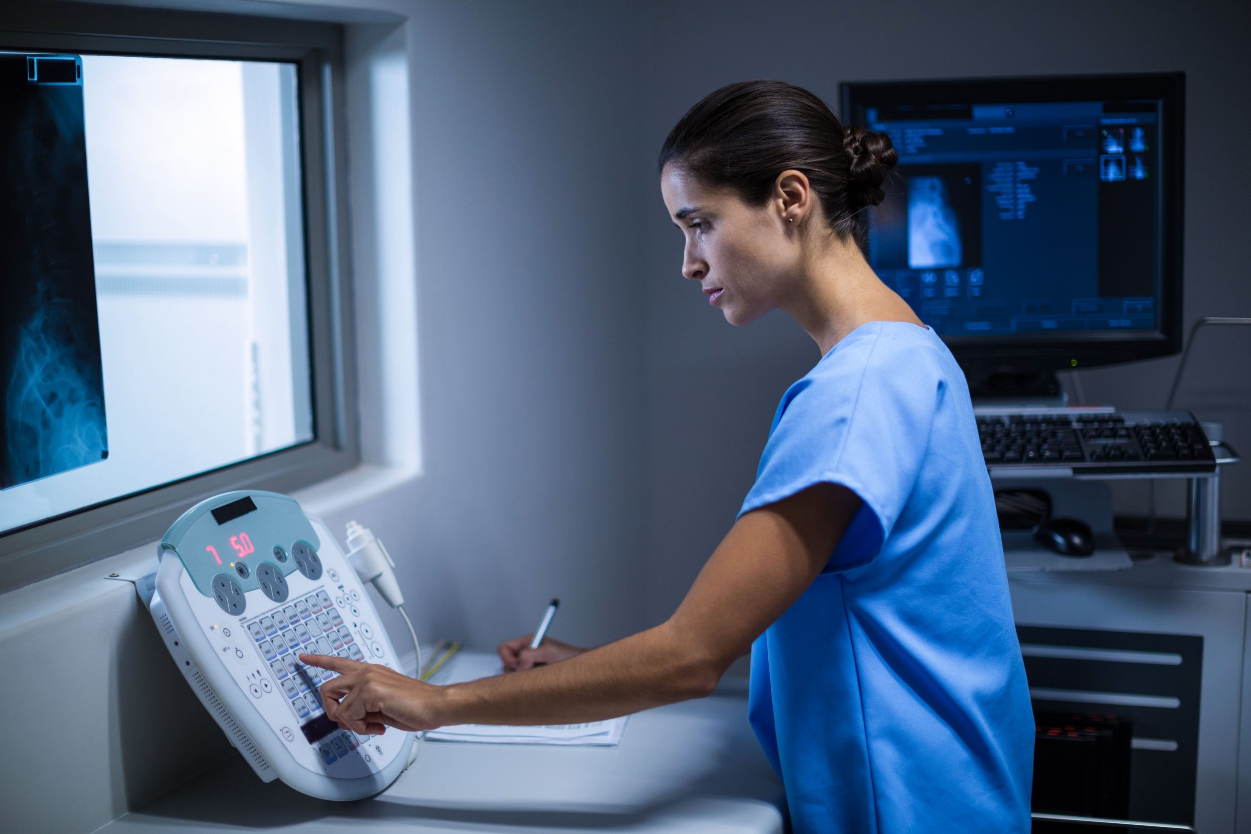
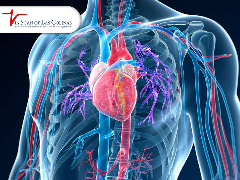
Gallbladder disease CT scan
Computed tomography, often known as a CT scan or CAT scan, is a non-invasive diagnostic imaging method that uses X-rays and computer technology to create horizontal or axial pictures of the body, often referred to as slices. An organ, muscle, fat, and bone structure may all be seen in great detail on a full body scan. Using computer technology and X-rays, a CT scan may provide pictures of your gallbladder, pancreas, and bile ducts. Gallstones and other problems, including infection and bile duct or gallbladder obstruction, can be seen on CT images. Nevertheless, gallstones that you could have might also be missed by CT scans. CT scan technology is getting better by the day. ViaScan facility provides mobile CT scan services for patients’ convenience.
Choose Our Preventive Wellness Body Scan
Early Detection Saves Lives!
-
- Accurate
- Quick Result
- Affordable

Conclusion
The majority of gallbladder disorders may be addressed surgically, either by excising the gallbladder entirely or by removing gallstones. Redirecting the bile ducts allows surgeons to bypass the gallbladder and go straight from the liver to the small intestine. Gallstones can be detected by ultrasound, or a more accurate measure is to conduct a CT scan. With ViaScan’s modern and high-tech technological scans and mobile CT scan services, taking preventative measures is easy. It is crucial to take care of your health before times run out. Saving you time, money, and worries.