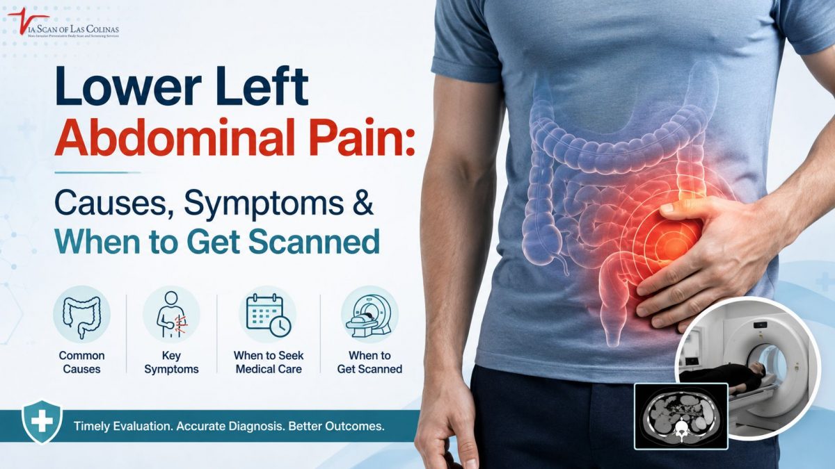
The lower left quadrant of the abdomen can be affected by a multitude of illnesses, from a flare-up of the gut to an emergency. It is where the descending colon, left kidney, left ureter and for females, left ovary and fallopian tube are located. The pain here is a message from your body to “pay attention”. This blog provides you with a step-by-step analysis of when to use imaging and warning signs and symptoms.
What are the most common causes of lower left abdominal pain?
The lower left abdominal pain is usually due to digestive, urinary, reproductive or musculoskeletal issues. The nature of the pain, its onset, and its accompanying symptoms give valuable hints to the involved system.
Gastroenterological, urinary, reproductive, musculoskeletal disorders.
- Diverticulitis — Inflammation of the pouches in the colon wall brings on sharp localized pain in the lower left quadrant of the abdomen, sometimes with fever, nausea and altered bowel movements, which are a warning sign of a digestive disorder that must be evaluated by a medical professional.
- Lower left pain radiating up to groin or back, often with urinary frequency or pain on urination and/or blood in urine, may be due to kidney stones or UTI (kidney infection) that are moving through the left ureter.
- For females, the left ovary may cause chronic/mild pain during their periods, or be the cause of sudden sharp pain that may be due to a left ovarian cyst, endometriosis or even ovarian torsion, all demanding varying degrees of medical urgency and evaluation.
- Physical effort or weakened abdominal wall, muscle strain or hernia — Musculoskeletal pain from physical exertion or a weak abdominal wall which gets worse with exercise or pressure but no digestive or urinary symptoms.
What could be the serious condition if the lower left abdominal pain has symptoms?
Some symptoms in combination with lower left abdominal pain indicate that there is a serious problem that requires immediate medical attention.
Warning Signs that should not be ignored:
- If there is fever with lower left pain, then it is likely that there is some infection or inflammation such as diverticulitis or a kidney infection which needs professional evaluation and should not be treated at home or delayed beyond a few hours.
- If left abdominal discomfort is accompanied by blood in the stool or urine, this is a sign that you are suffering from a digestive or urinary issue that needs medical imaging to determine the exact cause and rule out serious underlying problems.
- If the pain is sudden, severe and quickly getting worse, it could be a surgical emergency like a ruptured cyst, ovarian torsion or perforated bowel and care should be sought immediately without waiting for symptoms to improve.
- If the vomiting continues or gas is seen to be trapped and the infant does not defecate, this may indicate an intestinal blockage which should be investigated immediately and should not be ignored for longer than several hours.
When should you seek medical attention for lower left abdominal pain?
If pain is sudden, severe or worsening quickly, get emergency medical help right away. A scheduled appointment is appropriate when the discomfort is mild and improving and occurs intermittently. The following table will help you make a decision.
If any doubts, please get medical evaluations. Persistent or new or worsening lower left abdominal pain is a sign to seek medical attention.
Emergency Symptoms vs. Non-Urgent Discomfort
| Seek Emergency Care | Schedule an Appointment |
| Sudden or severe pain | Mild pain that comes and goes |
| Fever above 101°F | Dull ache without other symptoms |
| Blood in stool or urine | Bloating or gas-related discomfort |
| Rigid or tender abdomen | Pain that improves with rest |
| Dizziness or rapid pulse | Cyclical pain tied to menstrual cycle |
When Is Imaging Recommended for Lower Left Abdominal Pain?
Imaging is advised when there is no physical or laboratory finding to determine the etiology or when a serious condition must be quickly established or excluded.
What role do CT Scans and Other Tests Play in Finding the Cause?
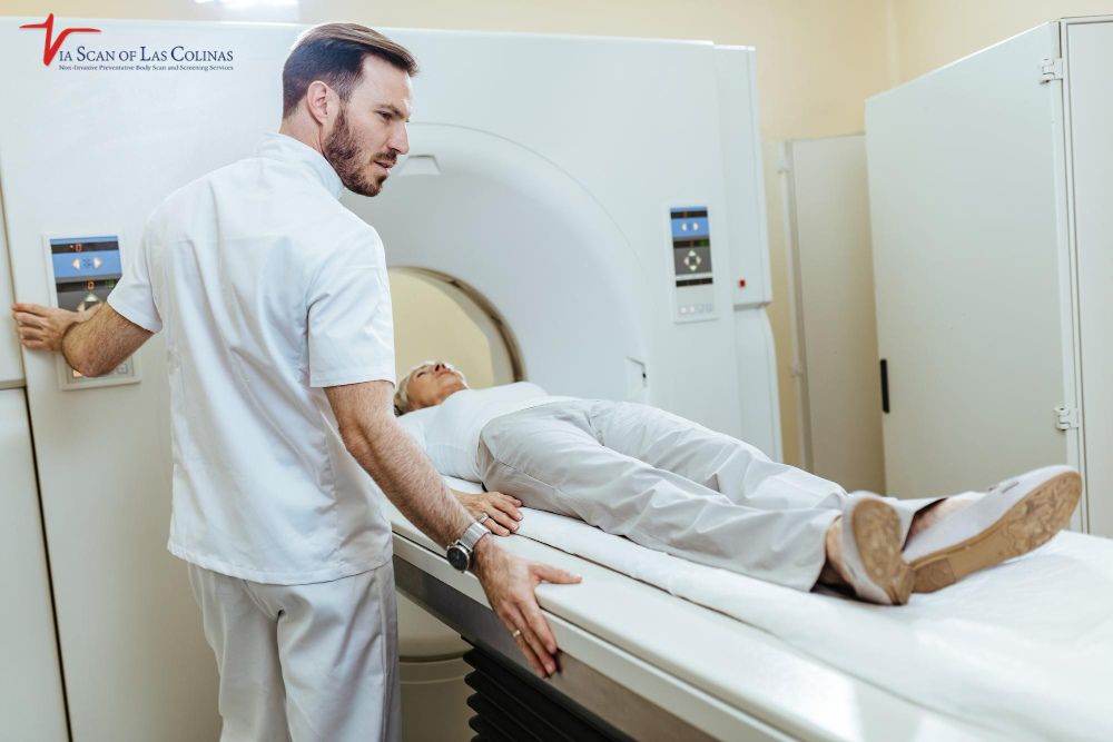
- A CT scan abdomen for lower left pain offers detailed cross-sectional imaging of abdominal organs, bowel, and surrounding tissue, and is one of the most informative and rapid tools available for the evaluation of the acute abdomen.
- A contrast CT scan abdomen and pelvis will show inflammation, blood vessels and boundaries between organs more clearly and is especially helpful to confirm masses that are not seen on other imaging, diverticulitis or kidney stones.
- Medical imaging for left abdominal discomfort that is chronic may include an MRI abdomen for chronic pain evaluation — preferred when soft tissue detail is required and repeated radiation exposure is a clinical consideration for the patient.
- When a woman has reproductive problems, ultrasound is usually the first procedure ordered before a contrast CT scan abdomen and pelvis or an MRI abdomen is ordered, and when the initial ultrasound results are not conclusive, a contrast CT scan abdomen and pelvis or MRI abdomen is ordered next to evaluate chronic pain.
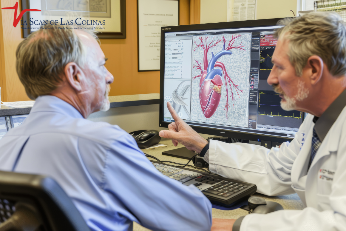
At ViaScan of Las Colinas, we provide a non-invasive preventive imaging service, which includes a whole-body scan that covers the abdomen thoroughly, for those who experience regular abdominal pain and are interested in a more comprehensive examination of what is going on inside. If there are cardiovascular issues in addition to abdominal symptoms, a heart scan can offer comprehensive information about the heart in a preventive evaluation. These are awareness tools, NOT emergency care, and are best used in union with instructions from a licensed health care provider.
Choose Our Preventive Heart Scan
Early Detection Saves Lives!
-
- Accurate
- Quick Result
- Affordable
Conclusion
Lower left abdominal pain is not something to take a stab at. The causes can be manageable or urgent; the appropriate action will depend on your symptoms. For anything new, persisting, or getting worse or anyone who wants to make proactive health choices, ViaScan of Las Colinas offer professional preventive imaging that can help with informed, early health choices.