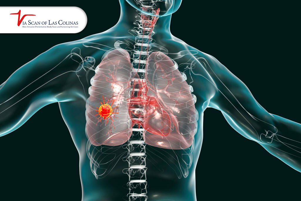
CT screening of lung cancer is one of the most essential steps you can take. You will need a CT scan for lung cancer in case you have been a previous or are still a current chain smoker, or you have other risk factors and symptoms of lung cancer. Lung cancer can be best treated at its early stages. Since lung cancer screening with a low-dose CT scan is possible, early detection becomes an important step to take before a single symptom is experienced. When to screen varies depending on your age, history of smoking, and personal risk factors. This blog takes you step by step on everything you should know about how the procedure is performed and what your findings imply in simple terms.
When should you consider getting a CT screening for lung cancer?
You are advised to get a CT screening for lung cancer, in particular, a low-dose CT scan, in case you belong to the group of people who are at a higher risk or have some significant factors.
The most popular guideline is that of the U.S. Preventive Services Task Force, who recommend screening in case:
- You are aged 50-80 years.
- You have a major smoking history (usually 20 pack-years or more)
- You have smoked or stopped smoking in the last 15 years.
You may want to talk to a doctor even when you do not fit within the standard guidelines, and you have persistent symptoms, including chronic cough, unexplained weight loss, chest pain, or shortness of breath. Although these symptoms do not necessarily indicate cancer, they can be a reason to conduct additional testing.
CT screening is aimed at the early detection of abnormalities before the symptoms manifest or the cancer develops. Lung cancer is much easier to treat when it is diagnosed at an early stage.
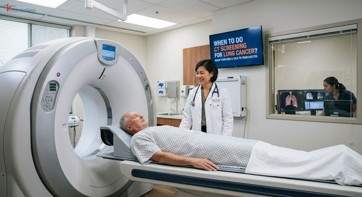
ViaScan of Las Colinas in Irving, TX, offers CT scanning service with advanced technology.
What is the duration that a CT scan takes for lung cancer?
The CT scan itself of lung cancer is a truly short process. The procedure is quick and straightforward for most patients.
Real Scan Time
The actual scan lasts about 10 minutes or less. But your overall visit with check-in, preparation and the scan lasts 30 to 60 minutes normally. Standard lung screening to identify cancer does not require any contrast dye, thus making it much easier, and less time-consuming to prepare than other forms of CT scans.
What Happens During the Procedure
- You come and check-in and fill in any necessary paperwork or health history forms.
- You put on a gown when necessary and get rid of any metal pieces such as jewelry.
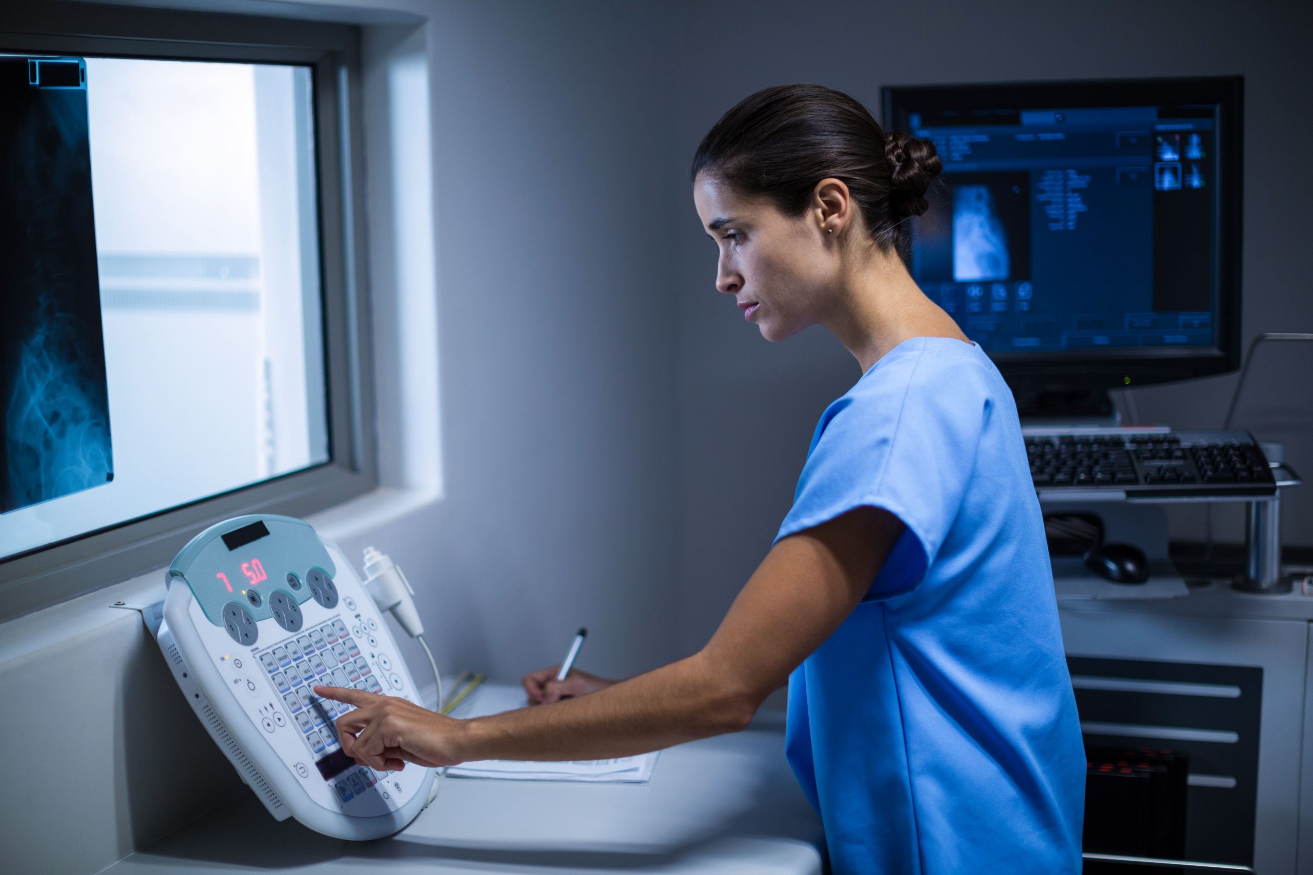
- You are lying on the CT scanner table, which moves slowly through the machine when imaging.
- A technologist will instruct you on a few breath-holding techniques – usually holding your breath 5-10 seconds when taking pictures.
- The table passes through the scanner and X-ray images are taken in various positions.
- The scan is finished and you are clear to go – it does not take any time to recover and you can go about regular life at once.
What Do You Need to Do to have a CT Lung Cancer Screening?
Prior to a lung screening to detect cancer, preparation is simple and will not cause much inconvenience. In most cases, a low dose CT scan of lung cancer does not involve any kind of sedation, contrast dye or any major dietary restrictions as compared to some medical procedures.
Pre-Scan Instructions and Restrictions.
- Wear loose, non-metal and underwire-free clothes. You might be requested to switch to a gown and in that case, light clothes ease the process.
- Take off jewelry, piercings, and metal accessories before coming to the appointment, since the metal objects disrupt the quality of CT imaging and have to be removed before the scans.
- Also, make the imaging center aware of any recent illnesses, surgeries, or major shifts in your health condition prior to your appointment as this may be pertinent to your interpretation of results.
- Always take any particular instructions you have been given by your ordering physician or the imaging center directly because each individual preparation may differ by a few details depending on the protocols used by the facility and your own health history.
What is the Time to Results of a Lung Cancer CT Screening?
The second question that most patients pose after undergoing a CT scan to determine lung cancer is the speed at which they will get the results. The time frame is usually quicker than what most individuals anticipate.
Result Processing Timeline
The majority of low-dose CT pulmonary screening findings are discussed and reported in one to two business days after the scan. Certain facilities give results in less than 24 hours. Depending on how your appointment was made, results are usually provided by your physician, patient portal, or even the imaging center itself.
Review of Results and sharing.
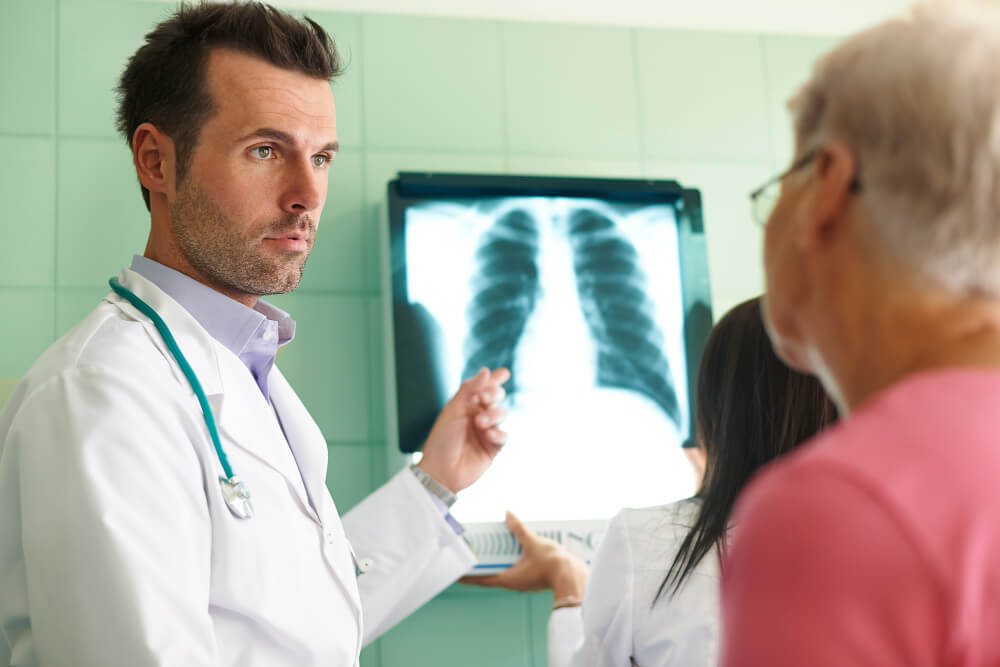
- A board-certified radiologist interprets the images of your CT scans to provide accurate results that can help your health provider to detect any abnormalities in lungs.
- A radiologist will issue a formal written report of all findings, such as the size, location, and character of any nodules or abnormalities found.
- The report is forwarded to you, which you can consult with your physician, who interprets the results in relation to your entire medical history.
- Your doctor calls you to talk about findings and next actions required – either regular annual check-ups or further testing.
In case you choose an imaging service from ViaScan of Las Colinas, our radiologist provides you with accurate, carefully reviewed results using the Lung-RADS system. The findings are reported in accordance with the standard reporting of the facility, and you are recommended to share the findings with your primary care provider.
What do the Results of your Lung Cancer CT Screening Mean to your health?
Getting your CT scan results of lung cancer can be a nerve-wracking experience but it is beneficial to know what your results actually mean in order to put things in perspective before you talk with your physician.
Normal vs. Abnormal Findings.
- Normal outcome is an outcome that indicates that no nodules or abnormalities were observed during the scan. This is a comforting result – but not an excuse to skip yearly screening provided you still fit within the recommended eligibility requirements. The screening for lung cancer is best done as a yearly process and not a one time thing.
- The majority of the abnormalities detected in CT scans of lung screening are benign nodules, small spots, which are common and usually harmless. The abnormal outcome is usually followed by further imaging or by a follow-up scan at the shorter interval to check any detected nodules to show how it will evolve, over time.
Follow-Up on the Results.
In case your results are within normal range, your doctor will advise on the next time to have your annual screening performed on the basis of your current risk profile. In case of nodules, follow-up recommendations will be done depending on the nodule size, nodule appearance and your risk factors. Big or suspicious nodules can lead to a referral to a pulmonologist or additional diagnostic tests. In every situation, your doctor acts as the guide to the following actions, and early diagnosis, even of something you need to keep an eye on, places you in the most advantageous situation in the future.
What is the Cost of a CT Scan to Screen Lung Cancer?
Price is a viable aspect to many patients, and knowing the price environment can allow you to strategize before booking your lung screening to check on cancer.
Pricing With and Without Insurance.
CT lung cancer screening is frequently no-charge as a preventive service to patients who have a meeting of Medicare or insurance eligibility criteria. When self-pay patients are not insured, the estimated price of a low-dose CT scan to diagnose lung cancer is between about $100 and $500, based on the facility and location.
Factors which influence the cost.
- Status of insurance covers and whether your plan covers the scan as an eligible preventive service on current eligibility rules.
- The nature of the facility – the hospital based imaging centers normally charge higher than independent outpatient imaging providers to the same scan.
- Geographic location, since imaging prices differ greatly by the region and metropolitan areas throughout the nation.
- The need to include extra services like physician consultation, follow-up imaging or referrals to a specialist according to your findings.
Who Can Have CT Lung Cancer Screening and When?
Not all people require a CT scan to diagnose lung cancer – it depends on certain age and smoking history requirements that are set by the major medical organizations and the U.S. Preventive Services Task Force.
Age, Risk Factors and Smoking History.
- The main candidates that are recommended to undergo annual lung screening to detect cancer are adults between the age of 50 to 80 years with a substantial smoking history according to the lung cancer screening guidelines.
- A considerable smoking history is considered to be 20 pack-years or more – it is the product of the number of packs per day smoked and the number of the smoking years.
- Smoking patients who fit the age and pack-year requirements are highly urged to talk to their doctor about annual CT scan as a lung cancer screening immediately.
- Quitters with less than 15 years since quitting and the age-pack-year requirements are also eligible to undergo screening – quitting smoking decreases the risk but does not eliminate it immediately.
Recommended Screening Guidelines
- Low dose CT lung screening should be performed annually, not a single scan. The ability to screen consistently over several years of time is what allows the early detection to occur.
- The eligible individuals should start screening at the age of 50 as per the current U.S. Preventive Services Task Force recommendations, revised as compared to the previous age of 55 years.
- Screening can be terminated in case a patient attains age of 80, has not smoked in over 15 years or has acquired a medical condition that is not likely to allow treatment of the detected lung cancer.
- Never start or stop any screening program without a licensed physician being consulted first, they do not always fit in the generalized guidelines and individual health circumstance will dictate how often or how often they can be used.
Choose Our Preventive Lung Scan
Early Detection Saves Lives!
-
- Accurate
- Quick Result
- Affordable
Conclusion
One of the most well-informed choices that you can make is a proactive approach to lung health, particularly having a history of smoking. ViaScan of Las Colinas in Irving TX is preventive imaging facility providing CT scan and whole-body wellness scans to help with early health awareness as an overall preventive care platform. These services are not a replacement for clinical diagnosis or physician directed treatment choices, nor do they substitute the medical care you already receive. Discuss with your medical practitioner the suitability of CT lung cancer screening. If you’re considering preventive screening, ViaScan offers fast, low-dose CT scans with radiologist-reviewed results helping you take an informed next step with your doctor.