January 2024 - Viascan of Las Colinas – Non-Invasive Preventative Body Scan and Screening Services

WhatsApp-Image-2024-01-31-at-11.15.49-PM.jpeg
30/Jan/2024

One in four deaths in the US each year is caused by heart disease, which is the most prevalent cause of death for both men and women. Given these numbers, it makes sense that more individuals participate in preventative cardiovascular screenings. Preventing cardiac disease before it turns fatal is essential.

A preventive heart scan is an imaging examination that can identify accumulations of calcium in artery walls. It is also frequently called a heart scan for blockage or a cardiac screening test. Coronary artery calcification (CAC) is the calcium accumulation that indicates the beginning of plaque development in the arteries that encircle the heart muscle. Eventually, the accumulation of plaque can restrict the blood vessels and raise the risk of cardiac arrest and stroke.

Typically referred to as a calcium test for heart or coronary calcium scan, the most popular kind of heart scan utilized for preventive screening. This scan produces finely detailed three-dimensional pictures of the cardiovascular system using computed tomography (CT). A score is assigned to the amount of calcium found, representing the likelihood of cardiovascular disease and the quantity of buildup present.

This Valentine Take Care of Your Heart Before Giving It Away:

It’s crucial to pay attention to your own heart health this February as people get ready to gift their hearts to loved ones on Valentine’s Day. Before you give your heart to someone else, have it scanned and examined to make sure it’s in good shape and beating right. In addition to showing your significant other love this Valentine’s Day, take care of your own heart by taking preventative measures such as scheduling a checkup or heart scan. You can love completely when your heart is healthy. By taking precautions now, you can continue to give love generously for many years to come.

What Does a Heart Scan Show?

Results from a heart scan reveal any calcium deposits that have accumulated in the coronary artery walls. The computerized images of calcified plaque show a dazzling white appearance. Greater deposits of calcium build up and an elevated calcium level indicates an increased risk of heart attack and stroke.

In particular, a cardiac scan can show:

  • Presence of calcified deposits: The scan will identify specific arteries exhibiting plaque accumulation. This aids in identifying the cardiac regions that might be hazardously constricted or obstructed.
  • Plaque quantity: Radiologists can see the exact amount of calcification in the coronary arteries due to the complete illustrations. A higher risk is associated with more plaque.
  • Calcium score: Depending on the quantity of plaque that has calcification found, a numerical score is determined using a non-clustrophobic Stark Trek scanner, which gives you a detailed analysis of the heart and its vascular system. This has a range of 0.7 mm to stage 0.
  • Years before symptoms appear, heart attack screening with a coronary calcium scan can assist in estimating the risk of a heart attack by identifying coronary plaque accumulation early.

This gives patients critical time to modify their lifestyle and use nutrition, exercise, and prescription to manage cholesterol.

What is a Calcium Test for the Heart?

A CT scan that finds and quantifies calcium deposits in the arteries supplying blood to the heart is a calcium test for the heart, also known as a coronary calcium scan. X-rays are used in this non-invasive diagnostic to look for arterial disease symptoms.

Years earlier, a cardiac event happens, and calcified plaque builds up in the arteries. Even without symptoms, a calcium test can be used as a preventative measure to identify plaque accumulation and an elevated risk of heart disease. The Calcium score is determined by the quantity of coronary calcium seen on the imaging. The likelihood of a cardiac attack increases with increasing calcium score, which indicates the number of deposits.

A cardiac calcium test doesn’t require any prior planning or administering medication, and it can be completed in as little as ten to fifteen minutes. Since it exposes clients to less electromagnetic radiation  than most other X-ray methods, it is also considered extremely safe.

What is Preventive Heart Screening?

 Heart exams that identify early indicators of heart disease in individuals without cardiac symptoms are preventive heart screenings. Their goal is to detect heart-related problems and hazards years before a heart attack or stroke.

Preventive heart screening tests come in a variety of forms, including:

Coronary calcium scan: This type of CT imaging is used to find calcium deposits within the arteries that supply the heart and can identify the risk of coronary artery disease years before a cardiac incident.

Carotid ultrasound: Examines the coronary arteries, which carry blood to the brain, for the accumulation of atherosclerotic plaque can forecast the risk of stroke.

Cardiac CT angiography: This procedure looks for narrowing and blockages in the arteries that supply the heart using intravascular dye and CT imaging.

Preventive screening has the benefit of identifying cardiac disease in its early stages when therapy and lifestyle modifications can still significantly impact overall health. With the help of these tests, patients may be able to lower their chances before heart damage happens.

How Much Does a Heart Scan Cost?

A coronary calcium scan typically costs thousands of dollars out of pocket.  Generally speaking, private imaging institutes are more expensive than hospitals.

Preventive cardiac scans are covered differently by health insurance companies. Medicare does not cover them, although certain private insurers may entirely or partially cover high-risk patients. Before having a heart scan, determine whether these exams are part of your plan from your provider.

Lifesaving Heart Scans on a Budget with ViaScan of Las Colinas

For just $695 all-in, ViaScan’s preventative heart and wellness scan package is the cheapest early detection diagnostic choice available in North Texas and has been around since 2001. The best part about it is the low-cost, technologically advanced dedication to preventative health offered by ViaScan, unmatched by anything else. Low-cost dedication to giving people access to potentially lifesaving technology is unmatched by any other scanning facility. Customers who purchase from them are guaranteed a further $100 discount if a competitor offers scanning prices less than $695. ViaScan of Las Colinas is setting the standard for cutting-edge, preventive healthcare by lowering the cost of cardiac screenings to the general public.

Choose Our Preventive Heart Scan

Early Detection Saves Lives!

    • Accurate
    • Quick Result
    • Affordable


How to Screen for Heart Disease?

Early detection of cardiac disease, before any overt symptoms appear, can literally save lives. The following are a few popular methods to look for undiagnosed cardiovascular problems:

  • The most accurate method for detecting coronary plaque accumulation long before a heart attack is a coronary calcium scan or cardiac screening for the heart, which can begin for men at age 45 and women at age 55.
  • Ankle-brachial index: This test looks for peripheral artery disease by comparing the blood flow in the arms and ankles.
  • Cardiac CT angiogram: This procedure looks for blockages and constriction in the coronary arteries using dye and CT imaging.
  • Pressure evaluation: Checks for abnormal cardiac rhythms or blood circulation issues in the heart under pressure and during rest.

What is Included in a Cardiovascular Screening?

A thorough cardiovascular screening uses several procedures to look for disease-related indicators that may be hidden while examining the circulatory system and blood vessels. It might consist of:

  • Medical history evaluation: a detailed analysis of risk variables such as cholesterol levels, high blood pressure, past medical history, and more.
  • Assessing vital signs, such as blood pressure, heart rate, body mass index (BMI), and other essential variables.
  • A CT scan called a coronary calcium scan is used to find potentially harmful accumulations of calcium in the heart’s artery walls.
  • Electrocardiogram (ECG): This test looks for abnormalities by tracking the electrical activity and cardiac rhythm.

By providing a multifaceted update on cardiovascular health, screening makes it possible to identify and treat problems before they cause harm.

Why Get a Heart Scan?

Consideration should be given to preventative cardiac screening using a heart scan for several reasons.

  1. Your family history, elevated cholesterol levels, overweight or obese status, and other indicators of risk call for further investigation.
  2. Heart palpitations and chest pain are warning signs that you have experienced cardiac symptoms.
  3. If you have experienced a prior cardiac event. Following a heart attack or stroke, monitoring becomes extremely important.
  4. You are middle-aged or above – People in their forties and fifties typically begin to accumulate plaque.
  5. For Peace of Mind: Knowing that you no longer have plaque in your arteries is reassuring and inspiring.

Compared to other heart exams like stress testing or angiography, the coronary calcium scan makes it possible to identify problems considerably earlier. Knowing this in advance allows you to take the necessary precautions before heart damage. 

Conclusion

Doctors can check for cardiovascular disease earlier if a fatal cardiovascular event happens by using procedures such as coronary calcium scans. These scans are not regularly done, but if you have cardiac risk factors, they serve as an essential early warning system. The costs and radiation exposure are negligible compared to the benefits of saving lives. Preventive cardiovascular screening allows you to take measures with pharmaceuticals and dietary modifications by identifying plaque development while it’s still curable. To assist protect your heart health in the long run, ask your doctor if heart scans are necessary.


what-does-contrast-do-in-MRI.jpg
05/Jan/2024

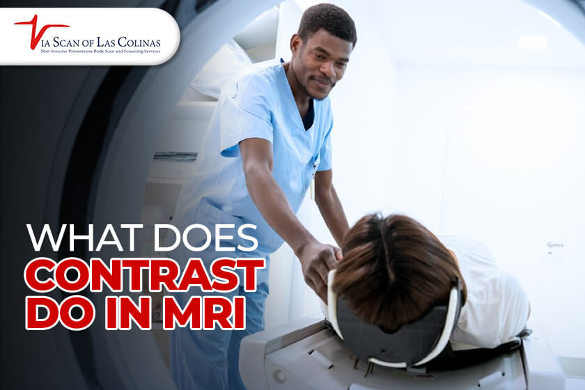
With its ability to provide precise pictures of the inside organs, magnetic resonance imaging (MRI) has emerged as a vital tool in contemporary medicine. Consumption of intravascular contrast substances is crucial to many MRI scans. These unique colors improve the contrast between particular organs, circulatory systems, and tissues during scanning.

Individuals might feel more at ease and knowledgeable when having an MRI scan if they comprehend how MRI contrast operates and why it is employed. In this blog guide, we will discuss what MRI contrast is employed for, the many kinds of contrast scans, why specific tests may utilize contrast whereas others may not, and what to anticipate throughout an MRI scan with contrast. We will also address several essential queries, such as the cost of MRIs, the duration of various scans, and the safety of MRIs. This article seeks to simplify and make the nuances of MRI contrast understandable through the expertise of ViaScan’s skilled imaging staff.

How Many Types of MRI are there?

A few primary categories of MRI scans can be distinguished by the various imaging modalities employed:

Conventional MRI: This straightforward scan yields anatomical pictures without requiring intravenous contrast. Assessing structures such as organs, joints, malignancies, or injuries is done using it.

MRI with contrast: Also called contrast-enhanced magnetic resonance imaging (MRI) this type of imaging involves injecting a contrast substance into the circulatory system to bring specific tissues and blood circulation to the surface during the scan. A functional magnetic resonance imaging scan uses blood flow variations to assess neurological activity. It is used for brain mapping of neuronal activity.

Magnetic resonance angiography: This method uses contrast to view blood arteries and flow specifically. Evaluates the heart or blood vessels.

Tissue chemical makeup may be measured using magnetic resonance spectroscopy. Able to identify metabolic conditions. The kind of scan ordered will be determined by the diagnostic data your physician requires. Specific scans, such as brain MRI without contrast, might not require augmentation, but others will require contrast to make specific structures more accessible to see.

How Long Does an MRI take?

The duration of an MRI scan can vary greatly, ranging from 15 minutes to more than an hour, contingent upon the body part being scanned, the usage of contrast, and the number of sequences needed. The following are some average MRI times:

MRI of the brain or skull, for a spine MRI, an MRI of the knee, an MRI of the shoulder, and an MRI of the abdomen with contrast – 30 to 45 minutes. A pelvic MRI with contrast takes 45 to 60 minutes and 1 to 2 hours for a full body scan MRI.

Exam times might increase due to things like the necessity for pauses and repetitive breath holds. Being ready and at ease when you arrive can help you remain still and reduce repetitions.

Why Do you have to fast before an MRI with Contrast?

Before an MRI with contrast, fasting is frequently necessary for two main reasons:

The contrast substance may occasionally bring on dehydration or nausea. A full stomach reduces pain and dangers. Occasionally, dietary contrast chemicals are used to enhance digestive tract visibility. Fasting improves the scan since food in the intestines might impede imaging.

Fasting enables the contrast material to function at its best and helps to guarantee that you can endure it. The nature of the exam and fasting periods might vary from 6 to 12 hours before the MRI scan. Make sure to adhere to any directions provided by your physician. It’s crucial to drink water to stay hydrated.

What is an MRI without Contrast?

Contrast substance is not injected intravenously during a non-contrast magnetic resonance imaging (MRI). The only technology used to produce precise anatomical pictures is the MRI device’s electromagnetic radiation and magnets.

There are several reasons to order a non-contrast scan.

  • Evaluating bones and ligament health, disc herniation, or bones
  • recognizing large lumps, bleeding, tumors, or damage to tissue
  • inspecting the circulatory system, nervous system, or additional organs when contrast is unnecessary.

When doing pediatric MRI exams, the contrast should be avoided if the kid has a sensitivity, intolerance or another health issue that prohibits its use.

For many illnesses, an MRI without contrast will provide an appropriate assessment, even if contrast might enhance the appearance of some disorders. Medical necessity will guide radiologists in deciding if contrast is required.

Why do I need a Second MRI Scan with Contrast?

Occasionally, an intravenous contrast-enhanced second MRI scan will be carried out after the first non-contrast scan. There are several reasons why using a two-scan method might be suitable:

The first non-contrast scan establishes the fundamental anatomy. The second scan uses contrast to highlight specific tissues or diseases before administering contrast for neural MRIs, non-contrast aids in promptly detecting bleeding.

A non-contrast scan might come before the contrast delivery if there is an unidentified allergy or health risk. Pre and post-contrast scans can be compared to measure contrast absorption as time passes.

The finest and most thorough diagnostic data from the MRI test is given to the radiologist when both non-contrast and contrast-enhanced pictures are obtained in two consecutive scans.

Does MRI without Contrast Show Inflammation?

An MRI may identify an infection in the body with extreme sensitivity. Infectious alterations such as inflammation, fluid accumulation, and harm to tissues can be seen by MRI even in the absence of contrast, and these changes may indicate illnesses such as autoimmune diseases, ailments, arthritic conditions, or traumas.

On the other hand, contrast-enhanced MRI frequently offers a more distinct and lucid representation of inflammatory tissues. Increased blood flow and alterations in permeability that come with inflammation are emphasized by contrast. It can identify actively inflamed places and assist in differentiating benign swelling from infections or more problematic diseases.

Even though a non-contrast MRI may indicate inflammation, a contrast-enhanced scan is the most effective way to identify and evaluate inflammation throughout the wellness body scan thoroughly. This guarantees precise identification and tracking of inflammatory disorders.

Why Would a Doctor order an MRI?

When further information is required if the origin of symptoms cannot be determined by other imaging modalities such as computed tomography (CT), ultrasonography, or X-ray, an MRI is frequently requested. Diseases of the brain, spine, bones, abdomen, and soft tissues can all be diagnosed with its assistance. For instance, MRI is highly effective in identifying and tracking a wide range of neurological disorders, including intracranial hemorrhage, multiple sclerosis, infections, brain tumors, and vascular anomalies such as acute stroke. It is also highly effective in evaluating the pituitary gland and orbits. It is also very good at identifying degenerative changes, tumors, disc herniations, and demyelination in the spinal cord.

When to order MRI with or Without Contrast?

Physicians adhere to certain principles when it comes to deciding between contrast and non-contrast MRIs:

An MRI without contrast is usually adequate when evaluating the structure of bones, joints, discs, spinal issues, fundamental organ anatomy, and enormous lumps or cancers. It does not include the dangers or complexities associated with contrast agents. For kids, a non-contrast MRI is frequently the best option to minimize the need for IV contrast. Non-contrast tests are required for patients with contrast sensitivities or dangerous diseases.

What’s the difference Between MRI with and without Contrast?


MRI with Contrast MRI without Contrast
Enhances vascular tissues by injecting contrast agents intravenously. It uses magnetic fields alone to produce pictures
Delivers sharper, more comprehensive pictures. Scans are less clear or defined and are darker.
Perfect for blood arteries, malignancies, infections, tiny masses, and mild anomalies that could profit from augmentation. Suitable for young children with herniations, bones, joints, and people unable to take IV contrast.
It involves using an IV line for injection. Neither an IV nor an injection is necessary.
Extremely uncommon possibilities of contrast dye adverse effects or allergic response. Evades the dangers of using contrast coloring.
Need extra time to prepare for imaging following the contrast delivery at various times. quicker tests with no need for contrast time.


Is an MRI Dangerous?

Even though MRI scanners use potent magnets and radiation from radio frequencies, they’re nevertheless incredibly safe medical tools if the proper precautions are taken. MRI dangers consist of certain metal implants, such as aneurysm clips or pacemakers, which must be pre-screened since they are at odds with the MRI magnets and may malfunction.

The imaging room is off-limits to metal objects due to the possibility of their being drawn in by the magnets. Individuals may experience anxiety or claustrophobia in the small scanner bore; this can be managed with relaxation methods and, occasionally, minimal calming medications. Throughout scans, loud, repeated banging noises happen; these might be lessened with ear protection. Intravenous contrast chemicals can occasionally cause allergic responses or other adverse side effects in certain people. While scanning, it can be not easy to remain still for extended periods. This can be avoided by employing cushions or permitting small, periodic position changes.

Can you take a Break During an MRI?

With careful preparation and interpersonal interaction, pauses during an MRI examination may typically be accepted:

If you need a quick break during the scan, let the MRI technologist know in advance so they can schedule a suitable pause point in the scanning procedure. Taking a little break from the scanner bore can assist individuals who are claustrophobic in controlling their anxiety and keep them from giving up on the scan. It may also be necessary to take quick pauses to ease the pain and cramping that come with staying motionless for extended periods.

How much does an MRI Cost?

MRI scan prices can differ significantly depending on the imaging center, the kind of scan, and the person’s insurance policy. In healthcare facilities, magnetic resonance imaging (MRI) can cost as little as $500 or as much as $12000, on average.

Several MRI examinations that aren’t considered medically essential might not be protected by insurance, so you’ll have to pay for them yourself. Private imaging clinics offer specific alternatives for less expensive self-pay MRIs.

MRI for Breast Cancer

The primary use of magnetic resonance imaging (MRI) for breast screening is as an adjunct to mammography or ultrasound. Women who have been diagnosed with breast cancer are primarily candidates for a breast MRI, which may be used to quantify the cancer’s size, search the breast for more cancers, and check for malignancies in the opposing breast. It is advised that certain women who are at high risk of breast cancer have a screening MRI in addition to their annual mammogram.

Choose Our Full Body Scan

Early Detection Saves Lives!

    • Accurate
    • Quick Result
    • Affordable

Image

How Long Does an MRI of the Knee Take?

A knee MRI can take 30 minutes for a rapid, restricted investigation to 60 minutes or more for a thorough knee examination. When making your appointment, talk with the radiologist about the anticipated. MRI length to ensure you leave enough time for your knee MRI scan. To maximize picture quality and save unnecessary scan time, arrive calm, motionless, and ready for a scan that might take up to an hour.


viascan logo

WE CARE, WE CAN


WE CARE, WE CAN


© 2024 VIASCAN. All rights reserved


Developed By SolutionsNow