What-Causes-Pulmonary-Embolism.jpg
21/Apr/2025

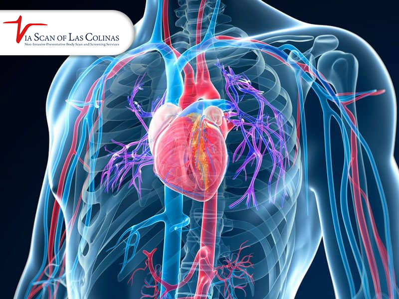
Slightly after you have been breathing normally, do you feel like you’re going to drown? Even if you’re on dry land. The pulmonary embolism makes its mostly unwelcome announcement with this terrifying sensation. A pulmonary embolism occurs when a blood clot travels through the veins and blocks one of the pulmonary arteries, which carry blood from the heart to the lungs. Each year, this dangerous condition affects thousands of Americans and can be fatal if not treated promptly. Allow us to explore this serious medical condition by answering some of the key questions about its causes and risk factors.

Why did Blood Clots travel to the Pulmonary Arteries?

Pulmonary embolism is most frequently caused by blood clots (which are simply clots of blood), formed in the deep veins of the legs (a condition called deep vein thrombosis, or DVT). These clots can break free and travel through the bloodstream to the lungs.

  • A blood clot’s journey takes place on a specific path.
  • Forms in a deep vein (usually in the legs)
  • Travelling through larger veins, it breaks loose.
  • Passes through the right side of the heart

It enters the pulmonary trunk (the main artery of the blood that passes through the heart to the lungs).

Small pulmonary arteries cannot accommodate their size and get stuck in them.

Since all blood passes through the lungs to pick up oxygen, all blood-borne clots will eventually end up in the pulmonary vessels unless they dissolve along the way. The pulmonary arteries branch so closely, creating trap points that clots can get wedged on the narrowing sections and stop blood flow to the tissue of some of this lung.

What Factors make a Patient at greater risk of having a Pulmonary Embolism?

Other things that can raise your risk of having blood clots that could develop into pulmonary embolism include:

Risk Category Specific Risk Factors
Medical Conditions • Cancer, Heart disease, COVID-19 infection and  Inflammatory bowel disease
Situational • Recent surgery or trauma, Extended bed rest, Long-distance travel 
Medications • Hormone replacement therapy and Birth control pills containing estrogen
Personal Factors • Pregnancy and postpartum period, Advanced age, Obesity and Smoking


People with cancer have a higher risk of blood clots, including pulmonary embolism.

How does a Blood clot impact the Pulmonary Trunk and lung function?

When a blood clot lodges in the pulmonary trunk or any of the pulmonary arteries, it begins a chain reaction of events.

  • Blocks blood flow to portions of the lungs
  • Prevents proper oxygen exchange
  • Increases pressure in pulmonary vessels
  • Works harder than normal, causing the right side of the heart to work harder.
  • In severe cases, it can lead to right heart failure.

However, the symptoms depend on the size and location of the clot. Small clots in peripheral pulmonary arteries may cause little, if any, symptoms, and large clots that block the pulmonary trunk would be immediately life-threatening.

Can a Pulmonary Embolism present before there are Symptoms?

Weirdly, many sufferers of pulmonary artery blockages often present with little or subtle symptoms, which are more likely to be confused with other things. Common symptoms include:

  • Sudden shortness of breath
  • Chest pain that gets worse when you breathe in deeply or cough
  • Rapid heart rate
  • Cough (sometimes with bloody sputum)
  • Feeling lightheaded or dizzy

Its silent nature makes it particularly dangerous because treatment delays compound mortality risk.

Why Early Screening for Pulmonary Embolism Can Be Life-Saving

Preventing the formation of blood clots is the best way to avoid pulmonary embolism. If you have risk factors, consider these preventive measures.

  • Do Not Sit Still – don’t sit for excessive periods
  • Stay hydrated, especially when travelling
  • Choose compression stockings for long flights

If prescribed, follow what your doctor recommends regarding blood thinners

Sudden shortness of breath, chest pain, coughing up blood or fainting can be signs of a pulmonary embolism. Call 911 or go to the nearest emergency department straight away. A pulmonary embolism is diagnosed in hospital, usually with a CT pulmonary angiogram.

Conclusion

When it comes to pulmonary embolism, don’t wait until the symptoms are severe; understand your risk factors and know your risk factors, ask your doctor about prevention if you are at higher risk, and get emergency help immediately if symptoms start


Why-would-a-doctor-order-a-chest-CT-scan.jpg
17/Mar/2025

Getting a recommendation from your doctor for requiring a chest CT scan generates both worries and uncertainty. Modern medicine uses this enhanced imaging technology to combine X-ray data measured from various directions to create detailed body section images. Your lungs, together with heart tissue and vascular structures and airways alongside chest bones, get shown in detailed 3D images through a CT scan despite basic chest X-rays providing only two-dimensional pictures. Advanced imaging systems let doctors recognize different medical issues that otherwise would stay undetectable and unclear. The reason for taking this test, as described by your doctor, will help reduce your concern and prepare you for the next steps. This piece examines the typical circumstances that trigger physicians to conduct chest CT scans in addition to explaining the scan findings and alternate imaging approaches and the essential patient requirements before the procedure.

What Medical Conditions Can a Chest CT Scan Diagnose?

The CT scan technology of the chest turns into an essential diagnostic instrument for healthcare practitioners to examine various thoracic region issues. A few millimeter lung nodules can be detected by CT scans, thereby enabling prompt diagnosis of early-stage lung cancer. Chest CT scanners prove highly effective in pneumonia diagnosis, particularly when standard X-rays produce unclear results to detect bronchiectasis, in addition to their ability to assess interstitial lung diseases leading to pulmonary fibrosis.

Coronary CT angiography is a heart scanning method used to evaluate coronary artery disease through analysis of heart blood vessels. It performs multiple diagnostics by showing vascular blockages in coronary arteries, measuring heart chamber dimensions and heart function, and checking the aorta status for aneurysms and dissections.

How Does a Chest CT Scan Compare to Other Imaging Options?

Three main factors, the suspected medical condition, the need for detail, and the degree of risk to patients from radiation exposure determine physicians’ choices for an imaging test. Laura Marie Fayad explains that there are differences in imaging tests. Understanding the distinctions between an MRI, CT scan, and X-ray will help you and your doctor decide which imaging modality is best for you. X-rays, CT scans, and MRIs are diagnostic procedures that let medical professionals view the body’s internal components. 

A standard chest X-ray shows overlapping structures in a single image, while CT produces cross-sectional images without overlap, giving more detail, for example of the area between the lungs (mediastinum). CT, however, uses more radiation than an X-ray.

What Should Patients Know Before Undergoing a Chest CT Scan?

Depending on the procedure selected and whether a specific chest CT scan includes contrast agents, there are different requirements for the chest CT scan. The main preparation step for patients who wish to get non-contrast scans is to remove only metal objects within the scan area. Patients must fast for some hours before contrast-enhanced studies and testing of kidney function might be done since contrast agents leak through the kidneys during elimination.

Iodine allergy needs to be notified to all healthcare providers by all patients, and patients must disclose their use of substances used in previous image procedures, too. Before experiencing treatment, previous reaction patients may be given premedication treatment containing antihistamines with steroids to minimize their chance of reaction. This is because radiation can harm developing fetuses, and patients need to come out about their pregnancy condition.

When Might a Doctor Order Follow-Up Chest CT Scans?

Follow-up imaging is an important central tool for assessing several chest conditions through specific time-based diagnostic-related timing protocols and clinical framework protocols used by healthcare providers. Regular CT scan imaging for cancer patients measures tumor changes such as dimensions and characteristics, and therefore, CT scan imaging determines treatment outcomes.
 After treatment, the medical team uses decreasing surveillance checks over the years in case there are signs of recurrence or metastasis. CT imaging, in combination, allows a medical practitioner to see any complications of thoracic surgeries and how the thoracic cavity processes healing following any thoracic surgery. Cardiac procedures require heart scan follow-ups to check the results of procedures and to diagnose stenosis following procedures, stent restenosis, or bypass graft failure.

Conclusion

In addition to detailed visualization of lungs, heart, and blood vessels as well as surrounding anatomical features, chest CT scans are also effective in diagnosing. ViaScan of Las Colinas offers coronary calcium scoring for adults without symptoms, which estimates future heart disease risk.  Their cardiac calcium scoring and coronary CT angiography programs involve the heart scanning capabilities of these programs, which can provide detailed results for detecting cardiovascular diseases at the initial stages and in the stage when treatments are proving most successful, just before the symptoms become evident.

References: U.S. Food and Drug Administration — What are the Radiation Risks from CT?

CardioSmart (American College of Cardiology) — Coronary Artery Calcium (CAC) Scoring


Cardiac-CT-Scan-vs-Angiogram-Whats-The-Difference.jpg
03/Mar/2025

Medical imaging helps to diagnose the conditions of the heart before those conditions become serious. The cardiac CT scan and CT angiogram are two currently used tests for evaluating the heart and blood vessels. So, how do these tests differ? Knowing their different roles can help patients decide if they should take steps to increase their heart health. Thus, this blog will break down the differences between these two procedures as to when and why each would be appropriate to use.

What Is a Cardiac CT scan?

A heart cardiogram imaging test, also known as a cardiac CT scan, is a specialized X-ray imaging test that enables detailed cross-sectional images of the heart and its surrounding structures. The procedure is noninvasive and helps doctors detect coronary artery disease, heart defects, and other abnormalities.

An X-ray beam captures multiple images of the patient lying on a table while that table moves through a CT scanner. The images are then scrambled to create an overall image of the heart. As a safer, more comfortable option for some patients, a cardiac CT scan does not require catheter insertion, as does a traditional angiogram.

What Is a Cardiac CT scan

What Is a CT Angiogram?

CT angiogram is a more detailed imaging test used to study the blood vessels, including the coronary arteries. CT scanning, in combination with a dye that’s absorbed into the bloodstream, highlights blockages or narrowing in the arteries.

It is especially useful for diagnosing conditions such as coronary artery disease, aneurysms, and blood clots. This contrast dye makes it easier for doctors to see potential problems with blood flow more clearly than is possible with a standard cardiac CT scan. CT angiograms have high accuracy in the diagnosis of arterial blockages, signifying a valuable tool for early diagnosis and treatment planning, as claimed by Smith et al. (2021).

How Do Cardiac CT scans and CT Angiograms Differ?

Both tests use CT imaging technology, but they are used for different purposes and procedures:

  • Objective: A cardiac CT scan is mainly used to evaluate the anatomy of the heart, including the heart chambers, valves, etc. A CT angiogram, on the other hand, focuses on blood flow and pollution of the arteries.
  • Contrast Dye: A cardiac CT scan will or will not use contrast dye, while a CT angiogram must use contrast dye to bring out the blood vessels.
  • Both tests are noninvasive. A CT angiogram requires intravenous contrast infusion, whereas a cardiac CT scan can be performed without dye in some cases.
  • Cardiac CT scan and CT angiogram diagnostic focus: A cardiac CT scan is used for general heart assessment, and a CT angiogram is used to detect specific arterial conditions.

Johnson & Lee (2020) also found that CT angiograms are more accurate in detecting the severity of arterial blockage, while cardiac CT scans are better for evaluating heart function and anatomy.

Which Test Is Right for You?

A cardiac CT scan or CT angiogram is chosen based on the symptoms and concerns the patient has. A cardiac CT scan can be recommended for patients with suspected structural heart problems or congenital heart defects or general heart screening by doctors. In contrast, a CT angiogram is often used in patients with chest pain, symptoms of coronary artery disease, or a high risk of heart attack.
You must consult with a healthcare professional if you are uncertain which test will be best for you. The latest technology is used to provide both “cardiac CT scans” and CT angiograms through ViaScan so that a correct diagnosis and better heart health can be provided.

Conclusion

Cardiac CT scans and CT angiograms are very useful diagnostic tools in determining heart health. However, they are not the same thing. A cardiac CT scan is a broad view of the heart’s structure, while a CT angiogram focuses more on detecting blood vessel abnormalities. That can inform patients and doctors on the best way for them to diagnose if there is something wrong with the heart. ViaScan offers advanced, noninvasive heart scans to help you stay on top of your cardiac health if you are looking for reliable cardiac imaging.


48130-1.jpg
26/Dec/2024

Breathlessness is a reason for much concern, and that is why searching for what is causing it most of the time is common. Chest X-ray is the most frequently used imaging modality to diagnose respiratory conditions. However, what does this imaging show, and how can it help to determine the source of breathing complications? In this blog post, the reader will learn how chest X-rays help in diagnosing shortness of breath, what chest X-rays are in the first place, what chest X-rays can reveal, and how a particular healthcare professional should read the results. We will also incorporate key research to provide a comprehensive view of this crucial tool for diagnosing clients.

Is Chest X-Ray Effective in Diagnosing Respiratory Issues?

A chest X-ray gives images of organs, tissues, and bones that are located in the chest, including the heart, lungs and arteries, so use can also check your lungs health by professional lung scan laboratory . It helps the doctors obtain a picture of these areas that may be the reason for shortness of breath, if any. Avoidant facets of Chess X-ray have been found useful and are only used in medical diagnosis of conditions such as pneumonia, pleural effusion, and heart failure. For instance, a cross-sectional descriptive study done in the year 2021 where patients suffering from acute respiratory distress had deformities of pulmonary edema were diagnosed using chest X-rays with the hope of enabling early occurrence of early treatments. The study was published in the Radiology Research and Practice by Smith et al (2021).

What Other Disease Could Be Diagnosed Through Chest X-Ray?

Several issues can be diagnosed with the use of a chest X-ray, including:

  • Cardiac Conditions

A chest X-ray can show the size and shape of the heart, signs of heart valve problems, fluid around the heart, and heart failure. It can also show deposits of calcium in the heart or blood vessels, which is possible evidence of a heart or blood vessel disorder.

  • Conditions of the lungs

An X-ray of the chest can reveal the status of the lungs, whether trapped air is around a lung, cancer, or infection. Furthermore, it may reveal other heart diseases, including congenital heart diseases, alongside chronic lung diseases like cystic fibrosis or emphysema.

  • States of the blood vessels

A chest X-ray demonstrates outlines of the big vessels next to the heart and can highlight aneurysms and other blood vessel problems.

  • Additional circumstances

A chest X-ray may also help diagnose tuberculosis, injuries to the rib cage, and medical device insertion. 

Chest X-ray is helpful in case of suspicion of respiratory problems, a harsh or persistent cough, chest injury, mechanical cardiac problems, or if the patient is having a fever. 

Is Chest X-ray a Promising Diagnostic Marker for Lung Diseases?

A chest X-ray is a useful first test. Like all these kinds of exams, they are excellent at demonstrating structural pathology, but some diseases may need further imaging or a biopsy. For instance, when a chest picture is taken to determine small lung nodules, initial symptoms of the disease or the early stage may not be diagnosed.

CT shows more detail than a chest X-ray and is better at finding problems such as early lung cancer or interstitial lung disease.

When is Chest X-ray helpful for shortness of breath?

If you experience persistent or severe shortness of breath, a chest X-ray may be warranted, particularly if accompanied by other symptoms like:

  • Accident, burn, injury, or sickness.
  • Leg swelling or fatigue may be signs of developing a heart problem.
  • Sudden severe shortness of breath — call 911, as it can be a sign of a blood clot in the lungs.

Healthcare providers generally use chest X-rays as the first diagnostic test to eliminate potentially fatal ailments before using sophisticated techniques. Given the multiple symptoms a patient indicates upon a physical examination, it is recommended that a general practitioner review all X-ray results.

Where Does Chest X-ray Stand in the  Plan of Differential Diagnoses?

Chest X-ray is not an isolated process but fits well within the imaging diagnosis process. Other tests may include blood oxygen measurement, lung function tests, CT or blood tests.

The frequency, timing, and outcomes of this condition vary by individual and clinical circumstances. Avoid a numerical estimate or timeline unless it is supported by a current, directly relevant medical source; discuss personal risk with a qualified healthcare professional.

Conclusion

Chest X-rays are also valuable in identifying probable causes of shortness of breath and giving information about lung and heart diseases. Starting from Heart Problems Visible on Chest X-rays to Tumor Detection in Chest X-rays, this diagnostic technique provides the basis for early and accurate diagnosis. Even though they may not give all the information necessary, they are useful in the preliminary detection of abnormalities and reference to other tests and procedures. Any person with such a condition must get to see a doctor as soon as possible.

If you have shortness of breath, see your doctor, who will decide whether you need a chest X-ray. ViaScan of Las Colinas does not perform chest X-rays.


Untitled-design-min.png
23/Dec/2024

Symptoms and imaging findings must be interpreted in context. A healthcare professional can evaluate the person’s medical history and examination and decide whether further testing is needed; no single symptom or scan finding establishes a diagnosis on its own. Signs of blockages, when detected early, can greatly minimize the risk of developing complications. Although self-diagnoses should never replace a doctor’s opinion and practice, some things people can observe at home and practices that they can perform may give them an idea of what might be possibly dangerous and, therefore, may call for a doctor.

What Are the Signs of A Heart Blockage?

Such blockages cause some diagnosable symptoms that are usually seen as early signs of a disease. A simple heart CT scan detects heart diseases at early stages. Pain or discomfort in the chest, described as angina pain, pressure, or tightness, is one of the most frequent signs. It may radiate to the shoulders, arms, neck, or jaw, causing discomfort. Another important sign of the disease is dyspnoea: this occurs during simple activities or even while walking or in a resting position. Vague and unexplained tiredness may also be a signal of heart problems because the flow of blood is restricted, and the organ fails to pump the blood efficiently.

Other signs, such as a high pulse rate, sweating, especially cold drops, light-headedness, or edema over the lower limbs, can also signify problems within the cardiovascular system. These symptoms can be diagnosed and addressed early, which could save a life. 

Can Technology Support You to Closely Track Heart Health in Your Own Space?

Today, medical technologies offer methods for people to monitor their heart conditions from the comfort of their homes. Wearable devices such as smartwatches with features like heart rate monitors and Electrocardiograms (ECGs) can detect abnormalities like Atrial Fibrillation, which may also indicate a blockage. Sphygmomanometers are also crucially important, as hypertension, a risk factor for occlusive arterial disease, is commonly present.

These cannot diagnose blocked arteries or blood vessels but provide a lot of information regarding the state of the heart and motivate people to seek the advice of professionals. For example, CT scans are essentially in clinical facilities to analyze the degree of coronary artery calcification and, therefore, the probable degree of blockage in coronary veins.

Which changes to make in Your lifestyle may assist in avoiding a blocked heart?

It is, therefore, only wise that people who want to avoid incidents such as heart blockages undergo various changes that will enable them to achieve heart-healthy lifestyles. A healthy diet plan should, therefore, incorporate fruits, vegetables, whole-grain foods, and lean sources of protein. Low cholesterol diets, through the elimination of foods pointing toward saturated fats, trans fats, and sodium, can make a huge difference in cholesterol and blood pressure, hence avoiding blockages.

Aerobic exercises, such as walking briskly, swimming, cycling, and others, should be done for at least 150 minutes per week to support heart health. Mindfulness, yoga, or meditation can also help avoid the dangerous effects of stress hormones on the heart. Last but not least, one must quit smoking because tobacco affects the arteries and aggravates plaque formation.

When should you Go for Medical Assistance?

As observed, those involving home care and daily changes are useful, but effective and accurate results can’t be obtained from home monitoring. Some of the symptoms that require the client to seek medical advice include chest pain, breathlessness, or irregular heartbeats that do not go away. If attention is not sought early, one is likely to suffer a heart attack.

ViaScan: Your Partner in Advanced Heart Diagnostics

If you need an all-inclusive diagnostic, you should turn to advanced diagnostic technologies such as ViaScan. CT colonography (virtual colonoscopy) can be a colorectal cancer screening option for some adults, but it cannot remove polyps or obtain tissue samples. If a suspicious finding is identified, follow-up optical colonoscopy may be necessary. Discuss the appropriate screening method and interval with a clinician. These harmless procedures provide exact information about the status of the heart and the cardiovascular system so patients can work on altering their lifestyles. For instance, their CT scans can be highly accurate in diagnosing the illness called coronary artery calcification or in evaluating risks linked to coronary veins as well.

Conclusion

Healthy heart care is a lifelong process that involves knowing a number of facts, monitoring health status, and medical checkups. Signs, home technology, and a general outlook remain crucial to addressing the problem at the first stage. However, in high-end precise diagnoses such as the ViaScan, the health of the cardiovascular system can be fully and accurately assessed, and this can enable doctors to provide early treatment for blockages. CT scans are useful in helping identify CAD (coronary artery calcium) and further evaluating the coronary veins, which are among the services that will safeguard your heart. Today, paying attention to your heart for healthier living is equivalent to having a healthy heart.
Reference: CardioSmart (American College of Cardiology) — Coronary Artery Calcium (CAC) Scoring


2151681057-1.jpg
13/Aug/2024

A heart scan can determine your risk for heart disease at an early stage. It is an excellent way to monitor your health if you are worried about the condition of your heart. More specifically, a coronary calcium scan is a non-invasive procedure that quantifies the calcium content in the coronary artery walls. This calcium level might serve as a marker for atherosclerosis or coronary artery disease (CAD). This blog discusses the significance of coronary calcium scans. You can also explore other information regarding heart scans.

Why Doesn’t Insurance Cover Coronary Calcium Scan?

This is one of the most common questions asked by several individuals. The most important factor is that coronary calcium scans are more often seen as preventative than diagnostic by many insurance providers. The test is commonly considered an optional procedure since its main purpose is to assess the risk of future heart disease rather than to identify a current issue. The significance of these symptoms or findings depends on the individual’s history, examination, and the condition being investigated. A qualified healthcare professional can recommend appropriate tests, explain their limitations, and determine the next steps. Avoid treating a screening scan as a definitive diagnosis. If you are worried about the cost, you can check with your insurance carrier to see whether or not they give any coverage or discounts for preventative heart health exams.

How Long Does a Heart CT Scan Take? 

It usually takes less than 10 minutes to actually scan the heart. However, it may take around 30 to 60 minutes for the whole session, which includes preparation and consultation after the scan. The patient lies on a table that slides into a computerized tomography (CT) scanner. This captures high-resolution pictures of the patient’s heart. The process is not painful and involves just a short period of time. It is basically an option that is valuable for those who have some busy schedules.

What is the Treatment for a High Calcium Score?

You are more likely to have coronary artery disease and a high risk of heart attack if your coronary calcium scan indicates a high calcium score. The criteria from the calcium score guidelines assist medical professionals in determining the most appropriate course of action for you. In most cases, treatment consists of making changes to one’s lifestyle as well as taking medicine to control risk factors. A review of typical remedies is presented here:

  • Lifestyle Changes:

Important actions include adopting a diet that is good for the heart. You can increase the amount of physical exercise you get, give up smoking, and learn to manage your stress.

  • Medications:

Doctors may recommend cholesterol-lowering medication such as a statin, and other medicines to control blood pressure or blood sugar. Whether aspirin is appropriate depends on your bleeding risk, so do not start it without talking to your doctor. [1]

  • Further Testing:

Further evaluation of the heart’s health may need the use of imaging techniques such as angiograms or stress testing.

It is vital to have regular follow-up appointments with your healthcare practitioner in order to assess your progress. You can also make any required modifications to the treatment plan that you have been following.

Why No Caffeine Before Calcium Scoring?

The findings of the heart scan may be affected by caffeine. This is because your heart rate and blood pressure may both rise as a consequence of caffeine use, which may have an impact on the outcomes of the scan. For the best assessment of your coronary calcium levels, it is ideal to maintain a state of complete cardiac relaxation. Follow the preparation instructions you are given when you book; you may be asked to avoid caffeine for a few hours before the scan.

Where to Get a Coronary Calcium Scan Near Me?

ViaScan of Las Colinas, 349 East Las Colinas Blvd, Suite C, Irving, TX 75039, offers coronary calcium scoring. Call (972) 739-2840, Monday to Friday, 8 AM–5 PM, to ask about pricing and appointments.

References:

[1] CardioSmart (American College of Cardiology) — Coronary Artery Calcium (CAC) Scoring — https://cardiosmart.org/cac. 


2147763846-1.jpg
01/Jul/2024

A cardiac computed tomography (CT) scan is a technique that produces high-quality, three-dimensional (3D) pictures of your heart, major arteries, and surrounding structures by using numerous X-ray beams at different angles. Cardiac CT makes use of cutting-edge CT technology to more clearly see your heart’s anatomy and related blood veins, either with or without intravenous contrast (dye). Your healthcare professional can get high-resolution, three-dimensional pictures of your heart and major vessels with multi-slice scanning. This blog will look deeply into CT scan images and if a chest CT scan shows heart problems.

What would a heart CT be able to show?

An evaluation of the heart and accompanying blood vessels is done with a non-invasive, painless heart CT scan to look for any artery blockages or narrowing, as well as any issues with the heart’s pumping action that could point to potential heart disease. In a CT scan, an IV is used to provide a dye, commonly referred to as contrast. This scan helps identify a variety of cardiac diseases by enabling us to track the blood flow via blood arteries and the heart.

A heart or chest CT scan shows heart problems, and your medical professional will have access to:

  • Arteries in the heart provide it with blood. 
  • Heart muscles, valves, and chambers. 
  • Veins in the lungs. 
  • Abdominal aorta, as well as the thoracic aorta.
  •  Pericardium (Sac that surrounds your heart).

 Can a CT scan detect heart problems?

Your doctor may recommend a CT scan of your chest if you are showing signs of heart disease. When diagnosing cardiac illness, a CT (computed tomography) scan is the recommended approach and is a highly common operation. Traditional metrics for risk factor levels include cholesterol and blood pressure. Researchers concurred that a CT scan would be a more accurate predictor of risk than other methods. A physician can use this to determine whether there are any health issues affecting the organ or the blood vessels around it. Chest CT scan shows heart problems, including:

  • Anomalies in the congenital heart 
  • Determining one’s heart attack risk 
  • Examining the fluid in the sac that surrounds the heart, measuring the heart’s pumping efficiency, and looking for signs of plaque accumulation and coronary artery constriction, which medical professionals refer to as coronary artery disease.  
  • Anatomy of the heart to look for indications of a heart attack, such as scarring on the heart muscle 
  • Identifying anomalies in the big boats 
  • Determining if a post-operative coronary artery bypass graft is still open

 Does a chest CT scan show heart blockages?

For best results, a heart blockage must be discovered early. There are several methods for identifying heart blockages. Start with a routine examination and let your doctor know that you’ve been experiencing cardiac problems. The complete heart and the blood arteries around it may be seen on a CT scan of the heart. It may also demonstrate artery-clogging calcium accumulation that obstructs blood flow. Physicians may refer to this as a heart CT scan for calcium scoring or coronary artery calcium scoring. Physicians may occasionally recommend coronary angiography, an invasive technique that involves injecting a contrast dye directly into the arteries to reveal any blockages.

 Can a CT scan show a heart attack?

A chest CT scan, also known as a heart screening or heart scan, is a medical procedure used to identify calcium deposits in the plaque of individuals suffering from heart disease. They are a useful tool for detecting atherosclerosis before symptoms appear. Studies have shown that a chest CT scan can show heart problems, and with early detection, preventative measures can be taken. You have more coronary atherosclerosis and more coronary calcium you have. This increases your risk of developing cardiovascular issues later on. It’s crucial to keep in mind that a CT scan cannot accurately forecast events like a heart attack since certain types of coronary disease do not manifest in this way. To get more precise images of the arteries in your heart, your doctor could also request a coronary CT angiography (CTA) in addition to a CT scan.

Can a CT scan detect blocked arteries?

CT scans have demonstrated their value as a diagnostic tool throughout time for evaluating arterial health, identifying heart blockages, and identifying other vascular health problems. The arteries supplying the heart with blood are examined with a CT scan. It creates cross-sectional pictures of the heart and blood arteries using X-rays. The CT scanner provides digital data by taking a succession of X-ray beam pictures around the heart. The data is processed by a computer to produce cross-sectional images, also known as heart slices. Sometimes, a contrast agent, an iodine-based dye, is injected into the circulation to improve the pictures.

Conclusion

Early detection and the findings of your chest CT scan help physicians to identify heart diseases and take preventative measures. The test’s outcomes will determine the precise recommendations. There might not be any modifications to your existing treatment regimen. Your doctor could, however, advise you to alter your diet and exercise regimen. Not only may leading a healthy lifestyle prevent heart disease, but it can also mitigate its symptoms. In certain instances, medical professionals advise doing further testing to validate the results of the CT scan. It’s now simpler than ever to take charge of your health thanks to Via Scan Of Las Colinas chest CT scans and calcium score testing provided in Las Colinas.


CT-Scan-Show-Heart-Blockage-1.jpg
20/Mar/2024

It’s critical to take care of our health as we age. This includes adopting a healthier diet, exercising, and using screenings and preventive medications that can identify health concerns before they worsen. One condition that may be detected early and treated to help avoid heart attacks, strokes, and other heart disease-related problems is blocked arteries. In this blog, we will explore how you can identify this important risk factor at an early stage. Can a CT scan reveal obstructive arteries? What you should know is as follows.

A Heart CT Scan: What is it?

There are two main types of heart CT. A coronary artery calcium (CAC) scan uses a low dose of radiation and no contrast to measure calcified plaque in the heart’s arteries; it estimates future risk but does not show whether an artery is blocked. CT coronary angiography (CTA) uses contrast dye injected through an IV to show the inside of the arteries and can show narrowing or blockages; it is usually ordered by a doctor to investigate symptoms.

When is a Heart CT Scan Necessary?

Your doctor decides which heart test is appropriate for you. Typical causes of cardiac CT needs include:

  • If you have chest discomfort and/or believe you may have coronary artery disease, a noninvasive method to find out if your pain is coming from coronary artery blockages or narrowing is cardiac CT angiography. 
  • Inconclusive stress test: If the information obtained from a stress test cannot definitively address your health concerns, your doctor may suggest a cardiac CT scan to check for coronary artery disease or other heart issues.
  • Existence of additional heart disease symptoms: Similar to chest pain, you may benefit from a cardiac CT scan to assess your heart for potential heart disease if you have shortness of breath, neck, jaw, back, or arm discomfort, among other symptoms.

How Can Blocked Arteries Be Found Using a CT Scan?

Since its inception, CT scanning technology has advanced significantly. Doctors may use CT scans to evaluate the amount of calcium accumulation in a patient’s arteries since the imaging is so comprehensive. Using a CT scanner, the calcium score test procedure entails:

  1. The machine’s electronic X-ray detectors pick up the X-rays after the scanner delivers them through the body. 
  2. After processing the data, the CT scanner produces cross-sectional pictures. 
  3. The heart and blood arteries are then shown in crisp 3D by reassembling these photos.
  4. Your doctor will use the pictures to assess your risk of heart attack or other heart-related problems. Your healthcare provider may suggest medication and lifestyle modifications if you have a high risk of developing heart disease to lower your risk of clogged arteries.

Guidelines for Getting Ready for a Heart CT Scan

You should do some things before a CT scan, which your doctor will go over with you. Until your physician instructs you differently, you should take your medicine as directed. Nevertheless, four hours before your treatment, you shouldn’t ingest coffee. There’s also a four-hour smoking ban that you must adhere to. The test findings will be impacted by the heart rate rise caused by caffeine and nicotine.

Heart CT Scan Results and Lifestyle Changes

Your CT coronary angiography pictures ought to be available soon after your test. You will probably be able to discuss the test findings with the healthcare physician who requested the test. If your test results indicate that you have or are at risk of heart disease, your provider might discuss treatment options. It’s always a good idea to adopt lifestyle modifications to assist protect the heart, regardless of the findings of a CT coronary angiography. Some heart-healthy lifestyle modifications are:

Regular exercise is important. It aids in weight control and lowers blood pressure, high cholesterol, and diabetes, all of which are risk factors for heart disease.

Consume nutritious meals. A heart-healthy diet low in sodium, cholesterol, and saturated fat and high in fruits, vegetables, and whole grains may all help manage blood pressure, cholesterol, and weight.

Give up smoking. One of the main risk factors for heart disease, particularly atherosclerosis, is smoking. The greatest strategy to lower your risk of heart disease and its consequences is to stop smoking. Consult your healthcare physician about smoking cessation techniques if you require assistance in stopping.

Control medical conditions. Take prescription drugs as prescribed if you have diabetes, high blood pressure, or high cholesterol. Find out from your doctor how often you require follow-up appointments.

Heart Scan And Calcium Scoring

One of the most significant variables affecting your general health and well-being is the condition of your heart. This is why timing is crucial when receiving the appropriate screening tests. The coronary calcium score test, sometimes called a calcium heart scan or calcium score, is one of the heart CT scan procedures that is becoming increasingly well-liked for evaluating heart health.

Using CT (computerized tomography) technology, coronary artery calcium scoring looks for accumulations of calcium in the coronary arteries, which feed the cardiac muscle with blood rich in oxygen. A medical condition known as atherosclerosis, in which plaque builds up on the wall of an artery, is indicated in these arteries by the accumulation of calcium. The resulting plaque gradually narrows and stiffens the coronary arteries, limiting blood flow. A heart attack may occur if the arteries are clogged.

The total score produced by the coronary calcium scan is determined by the degree of calcification within the coronary arteries. Higher scores are associated with more plaque accumulation and an increased risk of stroke and other cardiovascular diseases.

Conclusion

The arteries that supply the heart gradually narrow and solidify as time goes on. The secret to averting future unfavorable cardiac events is to catch it early and stop more calcium buildup. A heart CT scan can provide important information about your risk and state of heart health. To help you reach the best possible heart health, ViaScan of Las Colinas offers precise coronary calcium scoring and individualized guidance using our state-of-the-art CT equipment and specially trained radiologists. 

FAQ

How does a CT scan show heart blockage?

During the CT, a dye (also known as contrast) is administered through an IV during the scan. The dye shows blood flow through the heart’s arteries and helps doctors diagnose a range of heart conditions.

Cardiac CT uses advanced CT technology, with or without intravenous (IV) contrast (dye) to better visualize your heart structure and associated blood vessels. With multi-slice scanning, your healthcare provider can get high-resolution, 3D images of your heart  and great vessels.

How to check heart blockage at home?

Monitoring your heart health at home can include taking regular blood pressure readings, measuring your heart rate, and even performing simple tests like the stair test. However, it’s important to remember that these methods are not a substitute for professional medical advice. If you notice any changes or have concerns, it’s always best to consult with your doctor and get heart scan at reputable facility like 123bodyscan for accurate assessment.

[1] CardioSmart (American College of Cardiology) — Coronary Artery Calcium (CAC) Scoring

[2] Dartmouth Health — Should You Get a Calcium Score?


sign-of-heart-attack.jpg
01/Feb/2024

In the United States, cardiovascular disease is the top reason for death for both men and women. Heart disease claims the lives of tens of thousands of Americans annually, or one in every four fatalities. It can save lives to recognize the early symptoms and indications that may appear in the days and weeks that follow before a heart attack. This post will discuss six indicators that could point to a heart attack as soon as next month.

February is Heart Month – Heart attacks are preventable – we don’t have a treatment problem. We have an identification problem – doctors rely on tests that guess or only discover heart disease at very advanced stages; secondly, people wait until it’s too late – as an unexpected heart attack in a seemingly healthy person is frequently the first & only symptom indicative of a heart problem!

ViaScan’s advanced technology and early identification capabilities, you can pave the way for a long and healthy life – a winning strategy for healthy longevity. What’s more, ViaScan offers the most affordable preventive scan package in North Texas, combining life-saving preventive heart and body scans for early cancer detection at just $695 – a price that has remained unchanged since 2001. With ViaScan’s commitment to low-cost, high-tech preventive health, you won’t find a better deal anywhere else. If you find an advertised heart and body scan for less than $695, ViaScan guarantees to match the price and further reduce it by an additional $100. Take advantage of ViaScan’s low-cost guarantee and prioritize your health today.

How Long Can You Have Symptoms Before a Heart Attack?

Signs before a heart attack can indeed start to show up weeks or even months in advance of a heart attack. A calcium test for heart or a heart scan for blockage remains the best option for getting an insight into your heart’s health. Discomfort or pain in the chest is the most prevalent early warning indicator. This could have an aching, burning, or constricted feeling. Instead of being steady, the pain could come and go. Anywhere in the chest can experience it, and occasionally, it spreads to the arms, neck region, jawline, or spine. Chest pain should be assessed by a physician immediately if it lasts more than five minutes or disappears for a while before returning.

Many other minor symptoms can appear weeks or months before a heart attack, such as tiredness, shivering, indigestion, loss of breath, and nausea. Observe any new or aggravated symptoms and talk to your doctor about them. If a cardiac event does happen, the sooner it is treated, the better.

How Long Does Your Body Warn You Before a Heart Attack?

According to experts, your system may give signs for days or even up to a month before a cardiac event actually occurs. The length and intensity of the initial symptoms, however, differ greatly between individuals. Days or weeks before a heart attack, certain individuals endure excruciating chest pressure and agony that brings them to the hospital for treatment. Some may only have minor symptoms, such as exhaustion or loss of breath, which they blame on other factors like aging or being overweight.

It’s critical to identify early warning indicators and not ignore them. It is important to assess prodromal heart attack symptoms as soon as possible, regardless of how sporadic or mild they are. Once a heart attack actually happens, calling 911 or receiving immediate medical assistance if symptoms last longer than five minutes may prevent death or irreversible heart damage. Your doctor should be informed of any new or worrisome symptoms so they can look for any latent heart problems.

How Long Can a Woman Have Symptoms Before a Heart Attack?

According to studies, women are more inclined than males to exhibit early warning indicators in the days, weeks, or months prior to suffering a heart attack. For women, the most typical symptoms include anxiety, dyspepsia, shortness of breath, unusual weariness, and sleep difficulties. uncommonly signs of a heart attack such back pain, jaw discomfort, or nausea without sensations of pressure in the chest are also more common in women.

Women may have signs of coronary artery disease weeks in advance due to more subdued symptoms. A lot of the time, warning indicators are written off as stress, aging, or other medical issues. Women are also more likely to put off getting medical attention, which can worsen heart problems. It’s critical that women do not minimize symptoms that linger or flare up occasionally. Seek assistance if something seems strange.

What Happens Before a Heart Attack?

Deposits of fatty substances or calcium called plaque can burst within the blood vessels surrounding the heart in the few days preceding a heart attack. The resulting blood clot obstructs the cardiovascular muscle’s blood supply. The heart becomes damaged and weaker due to the limited blood supply. However, before a full-blown heart attack, the heart may convey warning signals about this occurring.

A heart attack could occur as follows:

  • Especially when exerting oneself, chest pain, pressure, tightness, hurting, or burning
  • Breathing problems and shortness of breath, particularly while active
  • arm, back, neck, or jaw pain that radiates.
  • light-headedness and nausea
  • exhaustion, lassitude, and dizziness
  • irregular pulse or palpitations in the heart
  • Sweating and clamminess are symptoms similar to the flu.
  • Stress or sleeplessness. 

Even if these symptoms seem minor, get medical attention immediately if any of them appear. It is advised to call the Emergency number if symptoms last longer than five minutes. Treating warning symptoms promptly can stop a heart attack or significantly lessen cardiac damage with a heart attack screening.


Conclusion

Knowing the warning signals of a heart attack weeks or even months in advance may save your life. The most typical symptoms of a blockage denying the heart oxygen are chest tightness and pain, but other symptoms include nausea, sweat, exhaustion, and shortness of breath. Women are more likely than males to notice early warning indicators. If a worrying symptom persists for over five minutes, immediately dial the emergency number immediately. Talk to your doctor about screening methods and risk factors. Early detection of symptoms allows doctors to reduce heart damage or stop heart attacks with drugs or other treatments. It is essential to pay attention to your body’s indicators as it can prevent you from joining the alarming statistics of coronary artery disease.

ViaScan provides heart scans for blockage detection and sophisticated cardiac screening testing. Plaque accumulation can be found with our coronary calcium scan and calcium test for heart disease before it bursts and results in a heart attack. Heart attack prevention and screening services from ViaScan are cutting-edge and offer vital information to prevent heart attacks before they happen. To learn more about our cardiac screening test alternatives and how we can assist you in taking charge of your heart health, contact us. 


WhatsApp-Image-2024-01-31-at-11.15.49-PM.jpeg
30/Jan/2024

Heart disease is a leading cause of death in the United States; according to the CDC, one person dies
every 34 seconds from cardiovascular disease [1]. Given these numbers, it makes sense that more individuals participate in preventative cardiovascular screenings. Preventing cardiac disease before it turns fatal is essential.

A preventive heart scan is an imaging examination that can identify accumulations of calcium in artery walls. It is also frequently called a heart scan for blockage or a cardiac screening test. Coronary artery calcium (CAC) is a marker of calcified plaque in the walls of the heart’s arteries. Any score above zero is evidence of coronary artery disease [2]. Eventually, the accumulation of plaque can restrict the blood vessels and raise the risk of cardiac arrest and stroke.

Typically referred to as a calcium test for heart or coronary calcium scan, the most popular kind of heart scan utilized for preventive screening. This scan produces finely detailed three-dimensional pictures of the cardiovascular system using computed tomography (CT). A score is assigned to the amount of calcium found, representing the likelihood of cardiovascular disease and the quantity of buildup present.

This Valentine Take Care of Your Heart Before Giving It Away:

It’s a good idea to pay attention to your own heart health this February, as people get ready to share their hearts with loved ones on Valentine’s Day. Heart disease can develop for years without obvious symptoms, so the best gift you can give yourself is to understand your risk. Start with a checkup with your doctor to review your blood pressure, cholesterol, blood sugar, weight, smoking and family history. If you are between 40 and 75, have no symptoms, and your heart risk is unclear, ask your doctor whether a coronary calcium scan could add useful information. Taking these steps now can help you keep sharing your love for many years to come.

What Does a Heart Scan Show?

Results from a heart scan reveal any calcium deposits that have accumulated in the coronary artery walls. The computerized images of calcified plaque show a dazzling white appearance. Greater deposits of calcium build up and an elevated calcium level indicates an increased risk of heart attack and stroke.

In particular, a cardiac scan can show:

  • Presence of calcified deposits: The scan will identify specific arteries exhibiting plaque accumulation. This aids in identifying the cardiac regions that might be hazardously constricted or obstructed.
  • Plaque quantity: Radiologists can see the exact amount of calcification in the coronary arteries due to the complete illustrations. A higher risk is associated with more plaque.
  • Calcium score: The amount of calcified plaque is converted into a number. A score of 0 means no
    calcified plaque was seen; higher scores mean more calcified plaque and a higher risk of a future heart
    attack. [3]
  • Years before symptoms appear, heart attack screening with a coronary calcium scan can assist in estimating the risk of a heart attack by identifying coronary plaque accumulation early.

This gives patients critical time to modify their lifestyle and use nutrition, exercise, and prescription to manage cholesterol.

What is a Calcium Test for the Heart?

A CT scan that finds and quantifies calcium deposits in the arteries supplying blood to the heart is a calcium test for the heart, also known as a coronary calcium scan. X-rays are used in this non-invasive diagnostic to look for arterial disease symptoms.

Calcified plaque can build up in the arteries for years before a heart attack happens., and calcified plaque builds up in the arteries. Even without symptoms, a calcium test can be used as a preventative measure to identify plaque accumulation and an elevated risk of heart disease. The Calcium score is determined by the quantity of coronary calcium seen on the imaging. The likelihood of a cardiac attack increases with increasing calcium score, which indicates the number of deposits.

A cardiac calcium test doesn’t require any prior planning or administering medication, and it can be completed in as little as ten to fifteen minutes. The scan uses a low dose of X-ray radiation and does not need an injection of contrast. [3][4]

What is Preventive Heart Screening?

 Heart exams that identify early indicators of heart disease in individuals without cardiac symptoms are preventive heart screenings. Their goal is to detect heart-related problems and hazards years before a heart attack or stroke.

Preventive heart screening tests come in a variety of forms, including:

Coronary calcium scan: This type of CT imaging is used to find calcium deposits within the arteries that supply the heart and can identify the risk of coronary artery disease years before a cardiac incident.

Carotid ultrasound: Uses sound waves to look for plaque in the carotid arteries in the neck, which carry
blood to the brain

Cardiac CT angiography: This procedure looks for narrowing and blockages in the arteries that supply the heart using intravascular dye and CT imaging.

Preventive screening has the benefit of identifying cardiac disease in its early stages when therapy and lifestyle modifications can still significantly impact overall health. With the help of these tests, patients may be able to lower their chances before heart damage happens.

How Much Does a Heart Scan Cost?

Self-pay prices for a coronary calcium scan vary by facility.

Preventive cardiac scans are covered differently by health insurance companies. Insurance coverage for calcium scoring varies and many plans do not cover it; check with your insurer before booking. Before having a heart scan, determine whether these exams are part of your plan from your provider.

Lifesaving Heart Scans on a Budget with ViaScan of Las Colinas

ViaScan of Las Colinas offers heart scan for cheap price, which includes [list]. The best part about it is the low-cost, technologically advanced dedication to preventative health offered by ViaScan, unmatched by anything else. Low-cost dedication to giving people access to potentially lifesaving technology is unmatched by any other scanning facility. Customers who purchase from them are guaranteed a further $100 discount if a competitor offers scanning prices less than $695. ViaScan of Las Colinas is setting the standard for cutting-edge, preventive healthcare by lowering the cost of cardiac screenings to the general public.


How to Screen for Heart Disease?

Early detection of cardiac disease, before any overt symptoms appear, can literally save lives. The following are a few popular methods to look for undiagnosed cardiovascular problems:

  • Coronary calcium scoring is most useful for adults aged 40 to 75 without symptoms whose 10-year
    heart risk is borderline, intermediate or uncertain, to help decide on preventive treatment such as
    statins. [3][2]
  • Ankle-brachial index: This test looks for peripheral artery disease by comparing the blood flow in the arms and ankles.
  • Cardiac CT angiogram: This procedure looks for blockages and constriction in the coronary arteries using dye and CT imaging.
  • Pressure evaluation: Checks for abnormal cardiac rhythms or blood circulation issues in the heart under pressure and during rest.

What is Included in a Cardiovascular Screening?

A thorough cardiovascular screening uses several procedures to look for disease-related indicators that may be hidden while examining the circulatory system and blood vessels. It might consist of:

  • Medical history evaluation: a detailed analysis of risk variables such as cholesterol levels, high blood pressure, past medical history, and more.
  • Assessing vital signs, such as blood pressure, heart rate, body mass index (BMI), and other essential variables.
  • A CT scan called a coronary calcium scan is used to find potentially harmful accumulations of calcium in the heart’s artery walls.
  • Electrocardiogram (ECG): This test looks for abnormalities by tracking the electrical activity and cardiac rhythm.

By providing a multifaceted update on cardiovascular health, screening makes it possible to identify and treat problems before they cause harm.

Why Get a Heart Scan?

Consideration should be given to preventative cardiac screening using a heart scan for several reasons.

  1. Your family history, elevated cholesterol levels, overweight or obese status, and other indicators of risk call for further investigation.
  2. If you have chest pain, shortness of breath, palpitations or other possible heart symptoms, see a doctor
    promptly, or call 911 in an emergency. A calcium score is meant for people without symptoms. [4]
  3. People who already have known coronary artery disease generally should not have a calcium scan,
    because it would not change their care. [5]
  4. You are middle-aged or above – People in their forties and fifties typically begin to accumulate plaque.
  5. A score of zero is reassuring and is linked with low short-term risk, but it does not rule out non-calcified
    (“soft”) plaque, especially in people who smoke or have diabetes. [2]

A calcium scan can show calcified plaque before it narrows an artery enough to cause symptoms. It does not show whether an artery is blocked. [3] Knowing this in advance allows you to take the necessary precautions before heart damage. 

Conclusion

For the right person, a coronary calcium scan adds useful information to a doctor’s risk assessment and can help guide decisions about lifestyle changes and medication. These scans are not regularly done, but if you have cardiac risk factors, they serve as an essential early warning system. The scan uses a small amount of radiation and is not recommended for everyone, so ask your doctor whether it makes sense for you. [3]  Preventive cardiovascular screening allows you to take measures with pharmaceuticals and dietary modifications by identifying plaque development while it’s still curable. To assist protect your heart health in the long run, ask your doctor if heart scans are necessary.

Sources
[1] Centers for Disease Control and Prevention — Heart Disease Facts.
https://www.cdc.gov/heart-disease/data/research/facts-stats/index.html

[2] Yale Medicine — Is a Coronary Artery Calcium Scan Right for You?
https://www.yalemedicine.org/news/is-a-coronary-artery-calcium-scan-right-for-you

[3] CardioSmart (American College of Cardiology) — Coronary Artery Calcium (CAC) Scoring.
https://cardiosmart.org/cac

[4] Dartmouth Health — Should You Get a Calcium Score?
https://www.dartmouth-health.org/articles/should-you-get-calcium-score

[5] Harvard Health Publishing — Do You Need a Calcium Scan?
https://www.health.harvard.edu/heart-health/do-you-need-a-calcium-scan