Weed-smokers-lungs-after-5-years.jpg
14/Jul/2025

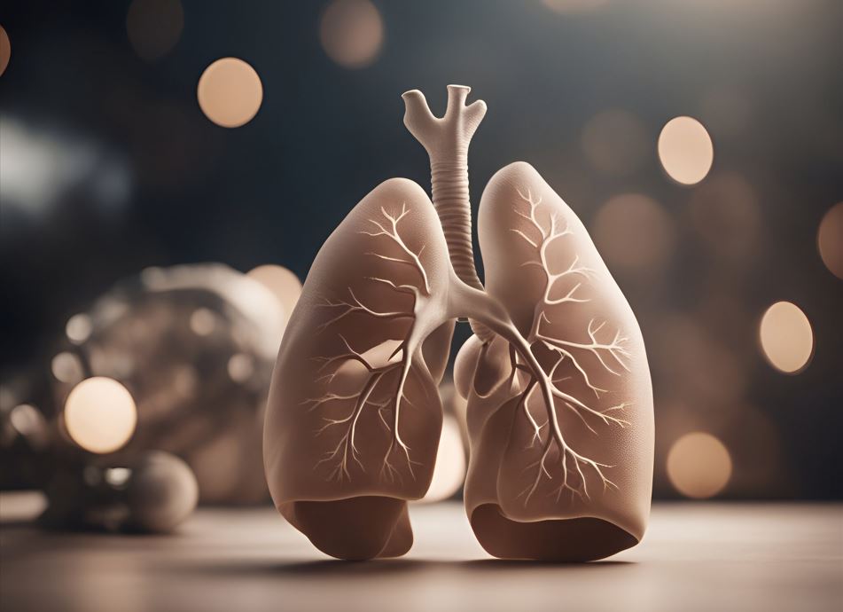
Your lungs are quite awesome! The lungs are in constant action all day and night, day in and day out, sucking air and giving your body the oxygen it requires. However, what if you smoked such weed over several years? It is one of the questions many people are interested in, and the answer is even more interesting than you can imagine.

Your lungs have experienced a major change after 5 years of smoking cannabis. They react just as any other organ in your body responds to what you feed it. Modern medical technology, such as a lung scan service, will enable you to know what happens to your chest. Being a radiology expert, I am going to tell you what happens to your lungs after several years of smoking cannabis. 

Does weed harm your lungs just like Tobacco?

And it is the question which people most likely ask, and it is a clever question! The answer to this is short: Both weed and tobacco are different to the lungs, but they involve inhaling smoke into the lungs.

Medical research says that cigarettes not only produce smoke but also more than 7000 chemicals in the smoke, and at least 70 of these chemicals are known to be carcinogenic. Nicotine is also present, which constricts the blood vessels and reduces oxygen circulation.

The cannabis smoke is rich in irritating chemicals like those of tobacco, yet it lacks the chemicals introduced during cigarette production. However, it is still smoke, and any smoke is likely to irritate your lung tissues.

However, here is the important part: both types of smoke can trigger irritation, cough, and alteration of your lung tissues. An early lung scan for smokers can detect if anything serious needs to be looked at.

The good news here is that lungs are tough. They possess remarkable cleaning mechanisms that actively remove particles and repair damaged components.

What Does the Lungs Change Five Years After Smoking Weed?

By your 5th year of smoking pot, your lungs have probably developed several ways to respond. Imagine that your lungs have been overworking themselves for the past 5 years.

Common changes are:

  • Heightened mucus production: your lungs produce more mucus. 
  • Airway irritation: The air sacs, which carry the air to your lungs, will be irritated and a little swollen. 
  • Lung tissue transformation: Some alterations may even be present in the small air sacs in your lungs (or alveoli).
  • Poor lung capacity: Others realise that they are not able to take in deep breaths.
  • Chronic cough: cough that persists.

What we must remember is that all these changes do not come up overnight. Your lungs have been slowly acclimating over the past five years. 

Can lungs repair when one gives up smoking weed?

And this is where the good news appears! The lungs are amazing curing engines. As soon as you quit smoking, they begin to mend themselves at work almost immediately.

This is the result of quitting:

In the first 24 hours, there is the commencement of the cleaning of your lungs of mucus and other foreign objects. It is possible that you will initially cough more as your lungs clear themselves.

  • During 1-2 weeks, the Increase in the size of your airway is reduced. 
  • In 1-3 months, your lung performance begins to increase. 
  • Within 6-12 months, you are often successfully treated for your chronic cough, and it improves significantly or disappears.
  • 1-2 years after: The large portion of irritation and inflammation in your lungs has healed. You are at a normal risk of lung infections again.

The earlier you quit, the better your lungs will be able to repair themselves.  So does stuff such as your general health, how much you smoked and whether you have other problems. Not every Weed Smoker Develops Lung Problems. Not all the people who smoke marijuana develop severe problems with their lungs! The same as not every person eating fast food after having heart disease, each person is different in the way they respond to cannabis smoke.

Those that influence your risk include:

  • The extent to which you smoke
  • The state of your general health
  • Your genetics
  • Your age

Numerous users of cannabis remain several years without getting notable complications in their lungs, particularly when they consume it in moderate proportions and keep their general condition in check. It is, however, advisable that you monitor your body and seek check-ups if you experience frequent coughing, feelings of breathlessness, and chest pains.

The Value of Early Detection and Professional Supervision

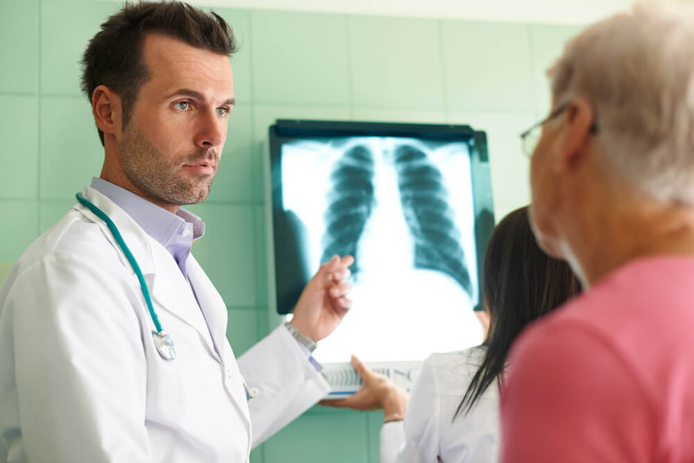
As a diagnostic imaging professional, I cannot overemphasise the importance of being aware of what is truly happening inside your lungs. Most changes in the lungs occur slowly, and you may not be aware of these symptoms until they have developed further.

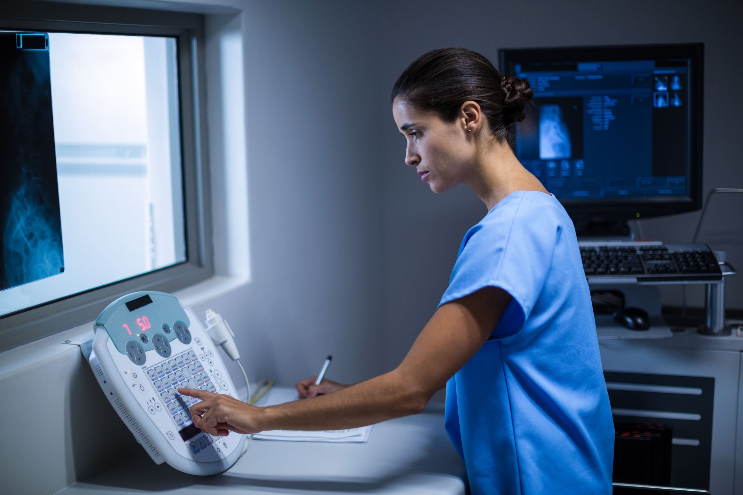
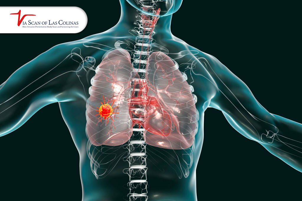
It is here that professional imaging services become so useful. A lung scan of a smoker: Your doctor can use a lung scan of some smokers to give detailed images of your lung tissues, so that she can find out areas of irritation, inflammation or even some changes that are not yet manifested or causing problems yet.

Sensitive imaging, like a CT scan of the lungs after smoking cannabis, has enabled the detection of tiny alterations in the lung tissues. These tests are painless and only take a few minutes, yet they may contain extremely insightful details about the state of your lungs.

Choose Our Lung Scan

Early Detection Saves Lives!

    • Accurate
    • Quick Result
    • Affordable

Conclusion

Lungs are wonderful organs that do an outstanding job of making you fit. They have already experienced a lot in the past 5 years using cannabis, and they are also amazingly able to heal themselves once they are provided with the opportunity. Using modern imaging technology at ViaScan, it has never been more convenient to check on how your lungs are functioning and monitor any changes.

Knowledge is power as far as your health is concerned. Regardless of whether you worry about your past cannabis use or need to keep track of your health condition in the future, professionally based services at ViaScan are the ones that can deliver much-needed, high-quality imaging and complete health tracking.


How-fast-does-a-blood-clot-travel-from-the-leg-to-the-lungs.jpg
07/Jul/2025

Blood clots are a common health concern for many people, particularly when they are exposed to news about clots spreading throughout the body and appearing in the lungs. The good news is that once you grasp how this is done, you can remain relaxed and be prepared in advance for what to look out for. You might think that it’s not very comforting, but with the facts, it’s not. At ViaScan, we help patients understand their risks and offer preventive lung and heart scans to detect potential issues early. There are no technical medical terms or clear facts that enable one to know what is going on with their body and when they must visit.

What is a blood clot? Where do they normally occur?

A blood clot is a patch that your body creates to stop the bleeding process. Just imagine it to be a bandage on your body. Blood clots are beneficial when they occur in the case of a cut, as they prevent excessive blood loss. However, in some cases, blood clots form in areas where they are not supposed to, such as in your blood vessels.

Which are the common locations of clotting?

The most usual places are the deep veins of your calves or thighs

  • Arms: Not very frequent, but possible
  • Pelvis: Occasionally, within the lower extremities of your pelvis
  • Other locations: Can develop in other body parts

When it occurs in a deep vein (it most often happens in your leg), you can be diagnosed with deep vein thrombosis or DVT. It’s like a livestock jam on your blood highway, which is unwarranted.

Why do the clots move to the Lungs?

Some of the reasons can be: 

  • Blood flow in your leg goes to the heart.
  • The blood flows into your lungs through your heart, which pumps it first.
  • The blood vessels in the lungs become smaller and smaller
  • The vein becomes too narrow, due to which the clot is stuck

A preventative lung scan can catch any blood clot that has travelled its way to your lungs. With clear imaging technology, you can take early action to improve your health. 

How Quickly Can a Blood Clot Travel from the Leg to the Lungs?

This is the question everybody would like to know! The trick is that you would be surprised by the answer. The rate varies according to several factors:

  • Clot size: The smaller ones move faster
  • Your heart rate: The quicker your heart rate, the quicker your travel
  • Blood flow: the quality of the blood circulation
  • Your activity level: Exercise influences blood circulation.

Important facts:

  • Not all leg clots make their way to the lungs
  • Some clots remain there and dissolve
  • Naturally, the body has a system to dissolve clots
  • Travelling can be prevented through rapid medical care

However, the most important thing to remember is that if you experience symptoms, do not wait to see if the clot travels or not; seek help directly.

What are the DVT early symptoms?

It is lifesaving to identify the early signs and symptoms of deep vein thrombosis. That is what to look out for:

General symptoms in your leg:

  • An enlarged leg compared to the other one
  • Pain: When walking, aching pain or cramping pain
  • Warmth: The area associated with the disorder feels hot when touched.
  • Redness: skin colour changes
  • Tenderness: it is painful to the touch

Important notes:

In most cases, only one leg is more involved than the other. Pain may manifest as a persistent cramp in a muscle that does not subside. If you experience them, particularly when they are sudden or occur more frequently, you should seek medical attention.

Which symptoms accompany a clot that has moved to the Lungs?

If a clot migrates to your lungs, it is referred to as a “pulmonary embolism,” and its symptoms differ substantially from those associated with leg clots.

Common signs Emergency signs
Shortness of breath: Suddenly, breathing becomes difficult Extreme chest pain
Chest pain Trouble breathing
Rapid pulse: Your pulse beats at a quicker-than-normal rate. Passing out
Coughing: There could be blood or pale, frothy foam brought up. Coughing blood
Leg symptoms: Can still experience pain or swelling in the legs

Is it possible to keep blood clots from moving?

There are numerous ways to avert the formation of blood clots and their movement:

Lifestyle prevention:

  • Keep moving:
  • Exercise: consistent physical movement aids healthy blood circulation
  • Stay hydrated by drinking lots of water.
  • Cease smoking: doing so elevates the risk of clots.
  • Maintain a healthy weight: Excessive weight puts pressure on veins.\

Medical prevention:

You may take medications that help prevent clots. Early detection combined with prompt treatment.  Health scans enable the detection of problems in the early stages.  Follow the movement recommendations provided by your physician.  Even minor movements help. Recognising the condition early is essential for averting serious complications. At this point, modern health screening services become crucial.

  • Ultrasound can detect blood clots in the legs.
  • CT scan: Shows any clots in the lungs
  • Whole-body scans: Thorough health screening
  • Lung scan: An imaging procedure dedicated to the lungs

Seek the professional assistance of qualified medical practitioners who will be able to determine the level of personal risk and advise accordingly on the necessary screening in relation to health history.

Choose Our Preventive Lung Scan

Early Detection Saves Lives!

    • Accurate
    • Quick Result
    • Affordable

Conclusion 

The description of blood clots moving from the leg to the lungs may be frightening, but understanding how it occurs can help you remain calm and know what to do. It is essential to detect symptoms early and seek help promptly. And it all comes down to the fact that your body is spectacular at healing itself, provided you support it properly. Most individuals who get blood clots fully recover after treatment. It is good not to overlook symptoms and seek help when necessary.

In situations where you are concerned about your risk of developing blood clots and wish to learn about preventive health screenings, ViaScan offers a comprehensive health screening package that includes advanced imaging scans and full-body scans. One of the best investments one can make is to take charge of their health through early detection and prevention.


How-to-remove-mucus-from-lungs-naturally-1200x800.jpg
30/Jun/2025

The lungs are an amazing organ that constantly works to enable you to live and breathe. They are great, like air filters that filter every breath you breathe. However, at times, they become excessive and lead to coughing or make breathing more difficult than normal.

Many patients approach me as a medical professional with the intention of learning how to naturally clear and comfort their lungs. Advanced CT scan technology can provide valuable insights into your lung health, helping your doctor monitor changes over time. There are natural ways that may support lung health and comfort. However, persistent issues should be evaluated by a healthcare provider.

In the same way we have today, natural remedies for that phlegm, which have been practised successfully over generations, we also have modern diagnostic equipment that allows us to keep our lungs healthy. Now, you’re interested in learning how to clear your lungs naturally and understanding the state of your respiratory system.

Why do we have mucus in our lungs?

It is not bad to have mucus in your lungs, but it is a defence mechanism of your body functioning as expected.

Consider mucus as a close protection of your lungs. Daily, you inhale a lot of such small particles as dust, pollen, bacteria, and others suspended in the atmosphere. To prevent unwanted guests from entering your respiratory system, your lungs secrete mucus to trap them.

On some occasions, however, your body produces additional mucus as:

  • You are suffering from a cold or a respiratory infection
  • You are exposed to irritants such as smoke or fumes of strong chemicals
  • You are allergic and have more mucus produced in your body
  • Bacteria or viruses have entered your body
  • You are dehydrated, and the mucus gets thicker

Is it possible to clear mucus in my lungs without medication?

The human body possesses natural healing powers, and there are numerous effective ways to support lung health without resorting to medication. Nevertheless, one should realise when the natural approach can be applied and when professional medical assessment is a necessary way out. 

However, natural methods may be highly effective but not sufficient in cases of serious conditions. The latest medical technology, such as CT scan imaging and other diagnostic procedures, can help identify underlying problems that require professional attention.

What are the most effective natural treatments to loosen lung mucus?

Some natural approaches have traditionally been used to support the body’s ability to clear mucus. Different practices have been applied over the generations without any danger and are supported by both ancient knowledge and the latest research on lung wellness.

  • Steam Inhalation: This is a natural method that is highly effective and quick. Warm, humid air breathed in also thins and loosens mucus, making it easier to cough up.
  • Saltwater Gargling: This is a simple technique that helps break up mucus in your throat and upper airways. Put half a teaspoon of salt in warm water and clean your throat.
  • Herbal Teas: Certain hot fluids help thin mucus and provide comfort.
  • Honey: Honey has traditionally been used to soothe the throat and may provide comfort for mild irritation. Raw honey, placed by the spoonful, can soothe and coat your throat.
  • Essential oil: You can open airways with eucalyptus and peppermint oil.

Keep in mind that these natural solutions are most effective when combined with enough hydration and sleep. 

What are the mucus-clearing foods?

Your respiratory system can be affected by certain medications and foods. The things you consume can either help your body clear lungs naturally or exacerbate the mucus buildup in your chest. I want to highlight the most nutritious foods that can help promote healthy mucus production and clearance.

Anti-Inflammatory Foods

  • Water is a must to take the target towards 8-10 glasses a day
  • Ginger is often included in diets due to its potential anti-inflammatory properties. 
  • Turmeric that aids the respiratory system
  • Garlic boosts the immune system
  • Leafy greens strengthen the entire lungs

Will Physical Movement Loosen Lungs’ Mucus?

One of the best natural methods to support your body in clearing mucus from the lungs and generally enhance breathing is physical movement. Movement can be described as the facilitation of your body to clear mucus more effectively.

  • Exercise also enhances the rate of breathing, and this facilitates the upward movement of mucus.
  • Workout enhances circulation, which supplies new oxygen to the lung tissues.
  • Chest Percussion: This involves tapping or vibrating movements on the chest and back to loosen mucus. A healthcare professional would be best suited to teach this.
  • Yoga and Stretching: Specific poses can stretch the chest muscles and improve breathing capacity.

One of the best ways to help your body clear your lungs naturally is to engage in regular, gentle movement. Nevertheless, in case of persistent respiratory issues, it might be advisable to have your lungs examined with the help of a non-invasive scan to see whether exercising is safe and reasonable in your case.

What is the Length of Time that Mucus Stays in the Body?

This is one of the most frequently asked questions, and the answer again will depend on several factors. Being aware of what to expect in terms of timing can make you realise when the natural remedies are taking effect, and you may require extra assistance.

  • In Minor Issues (such as allergies in the season or recovery from a cold):
  • It can help you improve in 24-48 hours
  • Many people may notice improvement within a few days, though timelines vary by individual health conditions.

The newer technology of diagnosis, such as CT scan imaging and other lung function tests, can be used to determine whether you have other underlying conditions that may be contributing to the delay in your recovery process. High-tech imaging of the lungs can reveal structural or functional problems that may be hindering your body’s ability to heal itself.

When and where should I visit a doctor for lung mucus?

It is the most significant question in the entire guide. Although natural methods may work quite well in the management of the mucus in chest conditions, it is important that you know when to consult professional medical help in the management of your health and safety.

Visit a Doctor as soon as you have:

  • Difficulty breathing or shortness of breath
  • Pain in the chest, particularly when it worsens or is severe
  • High temperature of more than 101°F that is prolonged
  • Bright red or blood traces in mucus

Where to go:

  • Your family physician is the first line of care
  • Emergency treatment on the same day to assess alarming symptoms

Have your lungs examined using a non-invasive CT scan, which helps identify problems that may otherwise remain unseen in standard X-rays and assists healthcare providers in making more precise diagnoses and treatment recommendations. Most health scanning services, such as ViaScan, have begun conducting health screenings that include lung imaging. Early assessment by a healthcare professional may help identify concerns before they progress, and this may help you use the most suitable and effective remedies under your circumstances.

Choose Our Lung Scan

Early Detection Saves Lives!

    • Accurate
    • Quick Result
    • Affordable

Conclusion 

Keep in mind that natural remedies for phlegm are more effective when used regularly; they are an integral part of a healthy lifestyle. Natural measures are great, but they work best when used in conjunction with informed healthcare choices. Lung health monitoring and assessment have become possible due to modern lung scan diagnostic technology which has provided us with unprecedented tools for assessing and monitoring lung health in a safe and non-invasive manner.

At ViaScan, we focus on your overall wellness by using wellness body scans and offer advanced imaging tools to support proactive health monitoring. We are up to date with the latest imaging technology, which enables us to produce clear images of lung health. This allows us to detect any issues at their earliest stage, monitor existing conditions, and provide you with peace of mind about your breathing health.


How-long-does-a-Colonoscopy-Take.jpg
23/Jun/2025

The question that many people ask is how long the colonoscopy procedure will take, how they will manage their work during the procedure, and when they can return to their normal life. As a healthcare provider, I often hear a lot of speculation and scary stories that many people have about colonoscopies.

The procedure of colonoscopy does not take as long as most people think it does. The primary process does not occupy as much time as a normal television program. However, the entire process consists of several stages, which are time-consuming. The following are the details.

What is a Colonoscopy?
A colonoscopy allows doctors to examine the inside of your large intestine using a small camera attached to a flexible tube. As a medical procedure, it utilises an endoscope, a flexible tube that is inserted into your body and features a lighted camera at its tip. Endoscopes come in various forms for different body sections. A colonoscopy involves the colonoscope entering your colon by your rectum and anus. It shows clear images of the interior of your big intestine on a screen.

What are The Benefits of Performing Colonoscopy?

Colonoscopy is an intensive examination of the large digestive tract (colon). The doctor operates using a thin, flexible tube with a small camera attached to it. That camera enables them to view the inner part of the colon on a television screen. It is as though a little explorer is being sent inside the bowels to take photographs and investigate issues.

The Reason Why People Undergo Colonoscopy.

  • Prevention of cancer. This is the most crucial reason. Small growths that develop before becoming cancerous are called polyps and can be detected using a colonoscopy. Searching for them early will literally save your life.
  • Health issues: When you experience pain in your stomach, changes in your bathroom habits, or other digestive problems, a colonoscopy can help determine the cause.
  • Routine examinations: After reaching a certain age, typically 45 or 50 years, doctors recommend regular colonoscopies to monitor the health of the colon.
  • Family history: If cancer is in your family, you may be required to have colonoscopies earlier or frequently.

The goal is to detect issues at an early stage.  They can be cured more easily by addressing minor problems before they escalate into major and costly ones.

How much time does a Colonoscopy really take?

The question everyone wants to know is how long it takes, which is between 15 and 45 minutes. In most cases, it is typically 20-30 minutes.

The reason behind the range?

  • Typical colonoscopy time: 20-30 minutes. This is what most people feel. The doctor spends enough time examining everything.
  • Complex colonoscopy: 30-45 min. Perhaps they need to clear a polyp, or your colon is winding and twisting, requiring more time to assess.
  • Very complicated procedures: It may take almost an hour, but this is rare and mostly occurs when numerous polyps need to be removed or when other complications are present.

The doctor will never rush your procedure. They strive to do a good job and ensure that everything is in order. Additional minutes of the process help avoid much more significant issues in the future.

Do I require sedation, and does that take time?

The majority of them do receive sedation, and yes, it does take some time from the total length of your visit. As an experienced doctor, I want to explain how this works and what it means for your schedule.

There are two types of Sedation:

  • Light Sedation: You will be calm and sleepy but not unconscious. It is the most widespread one. You are very likely to forget most of the procedure.
  • Deep Sedation: You will be in a deep sleep and will not recall any of the events. This can be particularly beneficial for individuals who are highly anxious or have previously undergone challenging procedures.
  • No Sedation: Some individuals opt to undergo the procedure without sedation. It can be done, although most individuals would prefer to be sedated, as it is much more comfortable.

The Effect of Sedation on Your Time:

  • Pre-procedure: The procedure will require you to wait up to 15 to 30 minutes to get sedated and to ensure that the medical team has you comfortable.
  • In the process, the sedation does not actually affect the duration of the actual colonoscopy.
  • Post-procedure: It will take you 30-60 minutes to wake up and ensure you are on your feet before you can leave.

In case of sedation, you will not be able to drive home yourself. You will have to have someone pick you up. This is one of the safety regulations that all medical facilities are adhering to.

What is the recovery time of a Colonoscopy?

The recovery time of a colonoscopy is almost always fast and painless! The post-procedure recovery phase, or the recovery time right after your procedure, usually takes 1-2 hours in case you received sedation. Throughout this period, you will be in a recovery room, spending between 30 and 60 minutes feeling somewhat groggy or sleepy. You may also experience some gas or bloating, which is normal, as the procedure uses air to assist the doctor in obtaining a clearer view. The medical team will examine your blood pressure and ensure that you are fine before releasing you to go home. To recover on the same day, most individuals are totally fine in 2-4 hours following the procedure. Most of the time, you can eat regular food immediately, but you may feel a bit tired due to the sedation.

How frequently will I need a Colonoscopy exam?

This likely is one of the most significant questions, and the answer lies in what the doctor finds during your colonoscopy. In case the results of your examination are normal, you will not have to take another colonoscopy at all until 10 full years. If you have small polyps during your procedure, you will likely require another colonoscopy in 3-5 years. Small polyps are quite common and are not particularly dangerous. The doctor simply wants to monitor the situation to ensure that things do not change. In case of a family history of colon cancer, you might require colonoscopies every 3-5 years rather than every 10 years. They may be required in 1-2 years in people with a personal history of inflammatory bowel disease.

Where to Get a Colonoscopy?

People are often confused, and it doesn’t have to be difficult to choose a place for a colonoscopy. You desire to have the trusted colonoscopy services, which are also affordable. The following are the key considerations.

Colonoscopy services are available in several kinds of facilities:

  • Gastroenterology hospitals
  • Outpatient surgery units
  • Full screening centres

A comprehensive screening centre like ViaScan can also offer packages that simultaneously screen multiple components of your health. That is convenient and smart. Other health screenings available at the best centres include wellness body scans, lung scans, heart scans, angiograms, and CT scans. This is a plus since you will be able to handle several screenings at a location, which saves you time and money.

What does a comprehensive scan include?

  • Full body wellness scans to identify any abnormalities discovery
  • Heart scans to detect any early symptoms of heart sickness
  • Scans of the lungs of individuals who smoke or those with breathing issues
  • CT images of a detailed view of areas
  • Angiograms to test the health of blood vessels

Having multiple screenings at the same time saves you the time of travelling to various medical institutions numerous times, i.e., time off work, time spent on the road, and, in many cases, it is more affordable to get the tests done in one visit than individually.

Choose Our Preventive Colonoscopy Scan

Early Detection Saves Lives!

    • Accurate
    • Quick Result
    • Affordable

Conclusion

Delaying this health screening is not worth it! The quicker you do it, the faster you can forget about it and move on with your good life.ViaScan offers top-notch colonoscopy procedures and a comprehensive range of other health checks. Their professional staff realises that you are a busy person and will do everything they can to ensure that your screening process is fast, comfortable, and complete.

ViaScan is dedicated to offering a range of screening services under one roof, allowing you to access your colonoscopy and other essential health checks, including a heart scan, lung scan, and comprehensive full-body wellness scan. This saves you time and helps you stay on track with your overall health. Keep in mind that a few hours of your day to have a colonoscopy may save you literally. And now that you understand precisely how long all of this will take, you can plan your day to the maximum and enter the whole process with confidence.


What-Causes-Pulmonary-Embolism.jpg
21/Apr/2025

Slightly after you have been breathing normally, do you feel like you’re going to drown? Even if you’re on dry land. The pulmonary embolism makes its mostly unwelcome announcement with this terrifying sensation. A pulmonary embolism occurs when a blood clot travels through the veins and blocks one of the arteries in the lungs that carry blood back to the heart. Each year, this dangerous condition affects thousands of Americans and can be fatal if not treated promptly. Allow us to explore this serious medical condition by answering some of the key questions about its causes and risk factors.

Why did Blood Clots travel to the Pulmonary Arteries?

Pulmonary embolism is most frequently caused by blood clots (which are simply clots of blood), formed in the deep veins of the legs (a condition called deep vein thrombosis, or DVT). Physical traits, such as these clots breaking free and travelling the bloodstream to the lungs, which they are prone to, are physical traits.

  • A blood clot’s journey takes place on a specific path.
  • Forms in a deep vein (usually in the legs)
  • Travelling through larger veins, it breaks loose.
  • Passes through the right side of the heart

It enters the pulmonary trunk (the main artery of the blood that passes through the heart to the lungs).

Small pulmonary arteries cannot accommodate their size and get stuck in them.

Since all blood passes through the lungs to pick up oxygen, all blood-borne clots will eventually end up in the pulmonary vessels unless they dissolve along the way. The pulmonary arteries branch so closely, creating trap points that clots can get wedged on the narrowing sections and stop blood flow to the tissue of some of this lung.

What Factors make a Patient at greater risk of having a Pulmonary Embolism?

Other things that can raise your risk of having blood clots that could develop into pulmonary embolism include:

Risk Category Specific Risk Factors
Medical Conditions • Cancer, Heart disease, COVID-19 infection and  Inflammatory bowel disease
Situational • Recent surgery or trauma, Extended bed rest, Long-distance travel 
Medications • Hormone replacement therapy and Birth control pills containing estrogen
Personal Factors • Pregnancy and postpartum period, Advanced age, Obesity and Smoking


A landmark study in the New England Journal of Medicine showed that cancer patients had an increased risk of developing venous thromboembolism (Venous thromboembolism includes pulmonary embolism) (Khorana et al., 2007).  

How does a Blood clot impact the Pulmonary Trunk and lung function?

When a blood clot lodges in the pulmonary trunk or any of the pulmonary arteries, it begins a chain reaction of events.

  • Blocks blood flow to portions of the lungs
  • Prevents proper oxygen exchange
  • Increases pressure in pulmonary vessels
  • Works harder than normal, causing the right side of the heart to work harder.
  • In severe cases, it can lead to right heart failure.

However, the symptoms depend on the size and location of the clot. Small clots in peripheral pulmonary arteries may cause little, if any, symptoms, and large clots that block the pulmonary trunk would be immediately life-threatening.

Can a Pulmonary Embolism present before there are Symptoms?

Weirdly, many sufferers of pulmonary artery blockages often present with little or subtle symptoms, which are more likely to be confused with other things. Common symptoms include:

  • Sudden shortness of breath
  • Pain that intensifies with forceful exhaling
  • Rapid heart rate
  • Cough (sometimes with bloody sputum)
  • Feeling lightheaded or dizzy

Nevertheless, it is estimated that up to 30–50% of pulmonary embolism cases are asymptomatic. Its silent nature makes it particularly dangerous because treatment delays compound mortality risk.

Why Early Screening for Pulmonary Embolism Can Be Life-Saving

Preventing the formation of blood clots is the best way to avoid pulmonary embolism. If you have risk factors, consider these preventive measures.

  • Do Not Sit Still – don’t sit for excessive periods
  • Stay hydrated, especially when travelling
  • Choose compression stockings for long flights

If prescribed, follow what your doctor recommends regarding blood thinners

Early detection is key to identifying concerning risk factors or symptoms. Viascan provides advanced imaging services that can help identify pulmonary embolism before it becomes life-threatening. Their latest CT pulmonary angiography can also detect even small clots in the pulmonary arteries, which can be addressed quickly and as soon as possible.

Choose Our Preventive Lung Scan

Early Detection Saves Lives!

    • Accurate
    • Quick Result
    • Affordable

Conclusion

Preventive screening is also offered via the Preventive Screen, which will help identify factors that are at risk of blood clots and cardiovascular disease so that you can take action to protect your health. All via the expertise of expert radiologists with convenient locations, this is how you know exactly where you stand when it comes to your pulmonary health status.

When it comes to pulmonary embolism, don’t wait until the symptoms are severe; understand your risk factors and seek appropriate screening early to save your life.


44a1c205-ec14-4928-8701-643681d0e751-1200x1145.jpeg
14/Apr/2025

Have you ever felt breathless at the very last stair while going up? Does breathing become a task for you? When breathing feels more like a task, your lung capacity and lung health might be in question. Whether you are interested in improving your climbing capability or just looking to improve your all-around wellness. Understanding how to increase lung capacity can change your picture for the greater part of your life.

In this article, we will discuss how you can increase your lung health. 

What Exactly Do We Mean by Lung Capacity?

In other words, lung capacity is the total amount of air you can breathe into your lungs. The average adult’s lungs can hold roughly 6L of air. However, with normal breathing, only a small percentage is used. Developing your respiratory system will allow you to use your natural capacity more, and depending on how much you appropriate, you could, over time, develop a greater capacity.

Is Regular Exercise Actually Able to Create Better Lungs?

Yes, regular cardiovascular exercise is one of the most effective ways to develop better lungs. Participating in physical activity that quickens your heartbeat. It deepens your breathing, strengthens your respiratory muscles, and makes them more effective at exchanging oxygen (Enright et al., 2006). The researchers found that the stronger the respiratory muscles are, the less energy it takes for them to function. Even when the muscles are resting, breathing more efficiently. 

Your weekly schedule should include these Physical activities:

Exercise through swimming delivers maximum advantages because it enables fitness training along with proper breath management.

  • Running or jogging
  • Cycling
  • High-intensity interval training
  • Rowing

Your respiratory function will improve significantly when you perform moderate exercise for just thirty minutes daily at least five times each week.

Do Breathing Techniques Provide Tips for Healthy Lungs Beyond Exercise?

Exercise establishes basic lung potential, but applying suitable breathing techniques will boost better lung capacity to higher levels.

  • When you use diaphragmatic breathing, you activate your diaphragm instead of shallow upper chest breathing.
  • Position your body on the floor while placing your hand on your chest, followed by resting the hand on your stomach.
  • Take deep breathing through your nose as your stomach expands without noticeable movement in your chest.
  • A slow exhalation through pursed lips helps your stomach to drop down.

What Environmental Factors Should I Control for Better Lung Health?

Your physical setting influences your breathing health to a significant degree.

  • A combination of proper air filtration units and sufficient indoor ventilation will help achieve clean breathing environments.
  • All types of smoke exposure must be completely avoided, as well as the inhalation of secondhand smoke.
  • Understanding which hazards exist from working with dust-related substances, chemicals, and other respiratory contaminants can help you better protect your lungs.
  • Check air quality reports in your locality while taking outdoor exercise during periods of poor air quality.
  • Keeping yourself hydrated enables your lung mucosa to function optimally.

Choose Our Lung Scan

Early Detection Saves Lives!

    • Accurate
    • Quick Result
    • Affordable

Preventive Screening helps Patients reach Superior Lung Health Levels

The basis of maintaining lung health consists of breathing exercises and physical activity. Together with preventive screening, it delivers extra reassurance to people. Especially to people who have risk factors for respiratory conditions. The advanced imaging technologies used by ViaScan during lung screening detect problems at early stages when no symptoms exist.

The low-dose CT technology at ViaScan performs quick, noninvasive lung assessments that give patients peace of mind. This provider’s prevention services provide customized risk assessments for smokers or those who used to smoke, people at occupational risk, and those with lung disease. Family members should use preventive screening from ViaScan to support their active respiratory wellness efforts.

Lung health benefits from your proactive lifestyle decisions combined with appropriate preventive screenings, which provide a complete form of lung care that helps you breathe better physically and emotionally for an extended period.


pulmonary-edema.jpg
07/Apr/2025

Can you visualize trying to breathe through a wet sponge? Each breath becomes a fight, air wadding slowly through water log passages. Patients who suffer from pulmonary edema feel this vivid sensation every day, which is a potentially fatal situation when fluid seeps into the sanctuary of your lungs. Pulmonary edema is a complex condition associated with heart failure that can also be caused by many other things but is so dangerous that it should be treated as soon as possible and correctly diagnosed, including with an advanced imaging test like a lung scan to determine if it is something other than lung cancer or pneumonia.

The Mystery of Fluid Where It Shouldn’t Be

Have you ever wondered how, in our lungs, the blood vessels do not make the organs wet with blood? Pulmonary edema is a fascinating balance between physics and biology when disrupted.

What exactly goes wrong when it all goes wrong?

Inside your lungs, there are millions of tiny air sacs (alveoli) where gas exchange miracle takes place. Those are the small air sacs where oxygen enters the blood and carbon dioxide leaves. Normally, these spaces stay dry and efficient in balance with forces that are very precisely right.

When pulmonary edema develops, the balance is shattered. 

  • First, it is for liquids to leak from pulmonary capillaries into surrounding tissues.
  • It then shoves off into the interstitial spaces between various structures.
  • It finally floods into the walls of the alveoli themselves.

That makes each breath a diminishing return, struggling to oxygenate as it becomes an increasingly impenetrable barrier to the gas. This can rapidly become life-threatening as if trying to extract oxygen from water rather than air. Pulmonary edema can evolve progressively over weeks to weeks or can develop quickly, in seconds, in hours. 

How Modern Imaging Uncovers the Truth?

How do doctors breach the enigma within your wrap of chest cavity? The story starts here. The humble chest X-ray remains the frontline detective but now has joined the investigation: A chest X-ray will reveal characteristic ‘butterfly pattern’ of fluid distribution; more sophisticated tools now come to the aid.

Studies in the European  Respiratory Journal (2018) by Assaad and colleagues suggest that the use of ultrasound can detect edema beforehand when it becomes symptomatic and can improve diagnostic accuracy by an impressive 17% in combination with clinical exam. Advanced images: CT scans help exclude pulmonary edema from mimics of the lung, such as lung cancer, pneumonia, or pulmonary fibrosis

How can you catch Edema Before a Crisis?

If you could only know that during times of crisis, your body speaks to you first, so to speak before the events actually come crashing down. It is the subtle signals that pulmonary edema sends, sometimes misread, sometimes dismissed, until the dramatic symptoms are present, that is the most fascinating aspect of pulmonary edema.

Consider these curious warning signs:

This includes unexplained weight gain due to secreted fluid retention.

  • Coughing occurs at night and almost completely disappears when you sit upright during nighttime.
  • Suddenly, you woke with the odd sensation that you were drowning
  • Sleeping with increasingly more elevated head positions

What is most interesting, however, is that many of these symptoms happen in patterns that can be recognized early on, sometimes before it becomes too late to prevent an emergency from occurring. Despite that, many people don’t trace those dots until that moment when someone is rushing to the emergency room.

What can be medical emergencies at high Altitudes?

In the nick of time, here’s a fascinating twist: perfectly healthy people can develop pulmonary edema just from getting too high too fast. It is different from other forms that plague people with medical conditions; it only strikes mountaineers and travelers who are at fault altitudes above 8,000 feet.

What makes this form unique? 

The condition triggers because of the very air, or, rather, the lack of it, around you.

  • The thin mountain air causes hypoxia (low oxygen).
  • It will spark a change in a blood vessel.
  • Pressure increases in the pulmonary circulation.
  • Where it shouldn’t, it starts to leak fluid.

Oddly, the progression begins with an out-of-the-ordinary breathlessness during activity, followed by never-ending coughing, gradual reduction of exercise capacity, and ultimately not being able to breathe even while you are standing still.

What can be the solution?

 The best medical intervention, even elegantly simple, sometimes happens: descent to lower elevation and supplemental oxygen.

Pulmonary edema is a fascinating meeting of cardiology, pulmonology, environmental and emergency care. Whatever the reason, the common theme is fluid accumulation, where there should only be air.

When outcomes depend on early detection through advanced imaging, the whole situation is transformed. Visualizing the exact pattern and distribution of fluids can tell doctors the difference between causes of the same symptoms that could require completely different treatments.

Controlling your respiratory health.

Are you having breathing difficulties that cannot be explained? Have you been visibly breathing any of the warning signs we have mentioned? Consider these steps:

  • Watch out for patterns of your breathing problems.
  • Look for anything to do with activity, time of day, or position.
  • Talk to your healthcare provider regarding even otherwise minor symptoms.
  • If these concerns continue to persist, don’t hesitate to ask a responsible provider about appropriate imaging.

As always, remember, knowledge about pulmonary edema is not only medical knowledge but potentially life-saving knowledge for bridging the perceptible gap between confusing symptoms and efficacious treatment.

Whatever the case may be, whether acute, like pulmonary edema, or chronic, comprehensive lung imaging can be useful to those who are worried about respiratory health. Modern non-invasive imaging technology provides the ability to see the details of the behavior of the lung tissue and helps in better diagnosis and better-targeted treatment.

Choose Our Lung Scan

Early Detection Saves Lives!

    • Accurate
    • Quick Result
    • Affordable

Conclusion

To understand pulmonary edema, one must understand the numerous causes and present various diagnostic approaches. ViaScan also provides lung scan services for those concerned with respiratory health. Their non-invasive imaging technology allows clinicians to visualize details of how lung tissue is behaving, so patients can be easily diagnosed based on the cause of their respiratory symptoms and their treatment plan clearly created. As both acute conditions like pulmonary edema and chronic diseases like lung cancer require early detection for the optimum outcome, ViaScan’s comprehensive screening services serve as an important facility in preserving respiratory health and recognizing and treating problems when they are not yet critical.


Chronic-Obstructive-Pulmonary-Disease-and-ICD-10.jpg
17/Feb/2025

Chronic Obstructive Pulmonary Disease (COPD) is a progressive lung disease occurring in millions of people globally. Persistent respiratory symptoms and airflow limitation are due to airway and alveolar abnormality. Normally, long-term exposure to harmful particles or gases is associated with COPD and usually to cigarette smoke. Medical coders, as well as healthcare providers, face the constant need to assign specific diagnostic COPD codes to maintain correct documentation and submission. This blog will give an insight into the significance of the ICD 10 coding system in identifying COPD conditions with an example of a popular ICD 10 code for COPD exacerbation.

How is COPD a Disease, and why does Accurately Diagnosing it matter?

COPD is a chronic respiratory disease characterized by two main symptoms: chronic bronchitis and emphysema. Breathing difficulties, chronic cough, and frequent respiratory infections accompany these conditions. Accurate diagnosis is very important as it helps in the appropriate treatment of COPD, which can avoid the patient’s readmission and improve their quality of life.

Lopez-Campos et al. (2016) argued that early diagnosis and intervention in COPD patients can significantly reduce mortality and healthcare costs. Thus, it is important to use the ICD-10 coding system correctly so that healthcare providers can accurately monitor or track the number of COPD patients, improve hospital patient outcomes, and facilitate insurance claims.

What is the ICD-10 code system, and how is that Applied to COPD?

The ICD-10 code used to classify and code health conditions is standardized worldwide. The ICD-10-specific codes for the stage and complications of COPD are available to help healthcare professionals be more detailed about the amount and type of the condition.

For example, the primary ICD-10 code ICD-9 for COPD without complications is J44.9, which refers to unspecified COPD. However, when patients’ symptoms worsen, or patients experience flare-ups, the code is J44.1, which stands for COPD, with acute exacerbation. The right use of the code, such as the ICD 10 code for COPD exacerbation, guarantees adequate care to patients and accurate reimbursement to healthcare facilities.

When and how is the ICD 10 Code for COPD Exacerbation used?

An exacerbation is a sudden increase in the severity of symptoms of COPD, such as increased breathlessness, excessive coughing, and wheezing that a doctor often treats. J44.1 is the appropriate ICD-10 code for COPD associated with an acute exacerbation. The absence of this code is critical, as, without it, you do not have met the criteria for routine COPD management; it also avoids hospitalization and emergency care for patients with severe respiratory distress.

Hurst et al. (2010) reported that COPD exacerbations are responsible for significant disease progression and deaths. Applying the ICD 10 code for COPD exacerbation not only indicates how severe the patient’s predicament is but also helps with making strategic treatment plans and better allocating resources in health facilities.

How Accurate ICD-10 Coding Benefits COPD?

ICD-10 coding is important and benefits patients and healthcare providers. These benefits include timely and appropriate care during exacerbations and accurate reporting of health data, which is crucial for public health research and resource planning.

Healthcare systems can track patterns in COPD exacerbations to enable better preventive strategies or fewer hospitalizations. Using the suitable ICD 10 code for COPD exacerbation guarantees healthcare experts legitimate recompense for their contributions, enabling them to succeed in attending to this.

Choose Our Lung Scan

Early Detection Saves Lives!

    • Accurate
    • Quick Result
    • Affordable

Conclusion

COPD is a very serious health condition that requires a correct diagnosis and proper medical coding to manage. During exacerbations, it is important that patients get the needed care, and the ICD 10 coding system helps a great deal. The research results underline the role of early intervention and adequate documentation in minimizing the complications of COPD.

ViaScan offers comprehensive diagnostic lung health screening and imaging solutions for those who need advanced diagnostic screening and services to help detect and monitor respiratory conditions such as COPD. You can trust ViaScan for accurate, professional health assessments that meet your needs.


4-symptoms-of-lung-cancer-that-you-should-be-aware-of.jpg
10/Feb/2025

Lung cancer ranks among the top cancer-causing deaths worldwide and is usually discovered only at an advanced stage. Detection at an early stage will improve survival rates, but such signs are often overlooked. Many of these symptoms should be recognized, and people should seek immediate medical evaluation, including a CT scan where possible, to make a big difference in outcomes.

Is a Persistent Cough a Painless Sign of Lung Cancer?

A persistent cough that haunts you for more than eight weeks is indeed an early indicator of lung cancer. Occasional coughing is not unusual, but a persistent cough that gets worse over time coughs up blood, or turns phlegm-colored should not be ignored. In this study by Rivera et al. (2020), one of the most common symptoms reported by lung cancer patients is persistent coughing, thought to be an external infection or allergies that last. If a cough doesn’t respond to usual treatments, it is a good idea to have a CT scan or, more specifically, a lung scan to find out what is causing it.

Why Does Shortness of Breath Occur in Lung Cancer Patients?

Dyspnea occurs when lung tumors block airways or fill fluid in the lungs. Gradually, this symptom may develop, and it may become apparent during physical activity. However, as it progresses, even basic tasks such as climbing the stairs or taking short walks can be very difficult. According to Wang et al. (2021), shortness of breath is commonly linked to reduced lung function resulting from the growth of tumors and, thus, a symptom for early detection. Seeking medical attention and having a lung scan to diagnose lung conditions can help if breathing difficulties persist.

Is Unexplained Weight Loss Related to Lung Cancer?

Unexplained weight loss is a common symptom of several cancers, including lung cancer. The spread of such cells to other parts of the body, when they create a different metabolism in the body, can cause rapid weight loss with no diet change. In addition to unintended weight loss, patients may have poor appetite or are extremely fatigued. Rivera et al. (2020) note that more than 60% of lung cancer patients at the time of diagnosis suffer considerable weight loss. A CT scan ought to be done if anyone has unexplained significant weight loss (more than 10 percent of body weight) within 6 months.

What Role Does Chest Pain Play in Lung Cancer Diagnosis?

Other alarming symptoms include chest pain. Tumor growth in lung cancer patients can cause chest pain if it presses on the chest wall, nerves, or ribs. It can be a persistent pain, or it can be intermittent and gets more painful with deep breathing, coughing, or laughing. Wang et al. (2021) suggest that chest pain stemming from the removal of lung tumors near the pleura (the membrane that surrounds the lungs) is more common. A person’s doctor can seek a CT scan to rule out any problems if they have unexplained chest discomfort, together with other symptoms such as coughing or breathlessness.

Choose Our Lung Scan

Early Detection Saves Lives!

    • Accurate
    • Quick Result
    • Affordable

Conclusion

The early warning signs of lung cancer (which, as you know, includes persistent cough, shortness of breath, unexplained weight loss, and chest pain), if recognized, will increase your chances of early disease diagnosis and success of treatment. However, with these symptoms, a lung scan to rule out or diagnose lung cancer in the early stages is an essential step. The advanced imaging solutions provided by ViaScan include precise CT scans that help detect lung abnormalities so the needed medical intervention can be done as early as possible. Act now to save your life by early detection.


48130-1.jpg
26/Dec/2024

Breathlessness is a reason for much concern, and that is why searching for what is causing it most of the time is common. Chest X-ray is the most frequently used imaging modality to diagnose respiratory conditions. However, what does this imaging show, and how can it help to determine the source of breathing complications? In this blog post, the reader will learn how chest X-rays help in diagnosing shortness of breath, what chest X-rays are in the first place, what chest X-rays can reveal, and how a particular healthcare professional should read the results. We will also incorporate key research to provide a comprehensive view of this crucial tool for diagnosing clients.

Is Chest X-Ray Effective in Diagnosing Respiratory Issues?

A chest X-ray gives images of organs, tissues, and bones that are located in the chest, including the heart, lungs and arteries, so use can also check your lungs health by professional lung scan laboratory . It helps the doctors obtain a picture of these areas that may be the reason for shortness of breath, if any. Avoidant facets of Chess X-ray have been found useful and are only used in medical diagnosis of conditions such as pneumonia, pleural effusion, and heart failure. For instance, a cross-sectional descriptive study done in the year 2021 where patients suffering from acute respiratory distress had deformities of pulmonary edema were diagnosed using chest X-rays with the hope of enabling early occurrence of early treatments. The study was published in the Radiology Research and Practice by Smith et al (2021).

What Other Disease Could Be Diagnosed Through Chest X-Ray?

Several issues can be diagnosed with the use of a chest X-ray, including:

  • Cardiac Conditions

A chest X-ray can show the size and shape of the heart, signs of heart valve problems, fluid around the heart, and heart failure. It can also show deposits of calcium in the heart or blood vessels, which is possible evidence of a heart or blood vessel disorder.

  • Conditions of the lungs

An X-ray of the chest can reveal the status of the lungs, whether trapped air is around a lung, cancer, or infection. Furthermore, it may reveal other heart diseases, including congenital heart diseases, alongside chronic lung diseases like cystic fibrosis or emphysema.

  • States of the blood vessels

A chest X-ray demonstrates outlines of the big vessels next to the heart and can highlight aneurysms and other blood vessel problems.

  • Additional circumstances

A chest X-ray may also help diagnose tuberculosis, injuries to the rib cage, and medical device insertion. 

Chest X-ray is helpful in case of suspicion of respiratory problems, a harsh or persistent cough, chest injury, mechanical cardiac problems, or if the patient is having a fever. 

Is Chest X-ray a Promising Diagnostic Marker for Lung Diseases?

Chest X-rays cannot be recommended, but they are nearly as accurate as they can get. Like all these kinds of exams, they are excellent at demonstrating structural pathology, but some diseases may need further imaging or a biopsy. For instance, when a chest picture is taken to determine small lung nodules, initial symptoms of the disease or the early stage may not be diagnosed.

A comparative cross-sectional study in The Lancet Respiratory Medicine established that X-rays are very specific, peaking at 70-80% in diagnosing lung pathology. Nevertheless, higher-caliber imaging provides superior biomechanical sensitivity and specificity, especially computed tomography, in diseases such as interstitial lung disease or early-stage lung cancer (Miller et al., 2020). This expands the applicability of Chest X-ray for Diagnosing Lung Conditions yet highlights the need for other tools.

When is Chest X-ray helpful for shortness of breath?

If you experience persistent or severe shortness of breath, a chest X-ray may be warranted, particularly if accompanied by other symptoms like:

  • Accident, burn, injury, or sickness.
  • Leg swelling or fatigue may be signs of developing a heart problem.
  • Sharp onset of dyspnoea (PE).

Healthcare providers generally use chest X-rays as the first diagnostic test to eliminate potentially fatal ailments before using sophisticated techniques. Given the multiple symptoms a patient indicates upon a physical examination, it is recommended that a general practitioner review all X-ray results.

Where Does Chest X-ray Stand in the  Plan of Differential Diagnoses?

Chest X-ray is not an isolated process but fits well within the imaging diagnosis process. Although ABG is relatively immediate and can be done noninvasively, other tests that may be mandatory include Pulmonary Function Tests, Computerized tomography, or blood tests.

A 2022 systematic review in the BMC Pulmonary Medicine found that chest X-ray augmentation of spirometry gave better performance in the diagnosis of chronic respiratory diseases than when imaging was used alone (Garcia et al., 2022). This supports extending the use of the Chest X-ray for Diagnosing Lung Conditions evaluation.

Conclusion

Chest X-rays are also valuable in identifying probable causes of shortness of breath and giving information about lung and heart diseases. Starting from Heart Problems Visible on Chest X-rays to Tumor Detection in Chest X-rays, this diagnostic technique provides the basis for early and accurate diagnosis. Even though they may not give all the information necessary, they are useful in the preliminary detection of abnormalities and reference to other tests and procedures. Any person with such a condition must get to see a doctor as soon as possible.

Choose Our Preventive Heart Scan

Early Detection Saves Lives!

    • Accurate
    • Quick Result
    • Affordable

 ViaScan offers comprehensive imaging solutions for different facets of your body, such as chest X-rays, so you and your healthcare provider can achieve the desired outcome. Please feel free to contact us today to learn more about our diagnostic products and how we can help you improve your health.