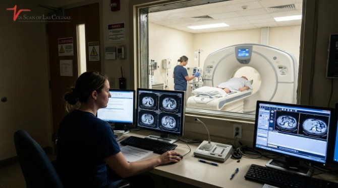
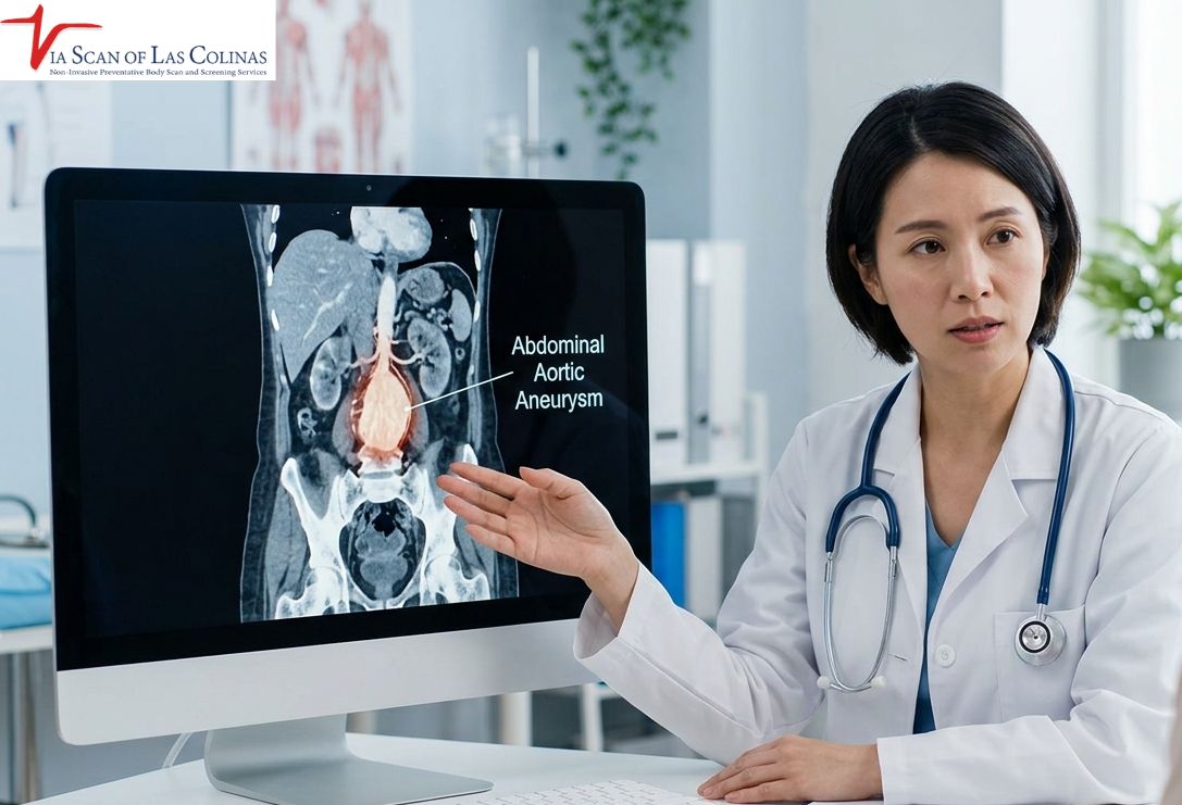
Aortic-Aneurysm-in-females-symptoms.jpg
24/Mar/2026

An aortic aneurysm is an abnormal enlargement or bulging of the aorta, the body’s largest artery. It can develop without noticeable symptoms, making early detection important, particularly in people with certain risk factors. Although abdominal aortic aneurysms are more common in men, women can also develop them and may experience serious complications. Understanding the possible symptoms, risk factors, and appropriate screening options can help women recognize when to seek medical attention. This article explains the warning signs of aortic aneurysms in females, how the condition is diagnosed, and when medical evaluation may be appropriate [1][2].

It is important to distinguish between an abdominal aortic aneurysm (AAA), which develops in the abdomen, and a thoracic aortic aneurysm, which develops in the chest. Their symptoms and recommended screening approaches can differ.

What are typical symptoms of an aortic aneurysm in females that can not be overlooked?

Many aortic aneurysms develop slowly and cause no noticeable symptoms, even as they increase in size. When symptoms occur, they depend on the aneurysm’s location and whether it is affecting nearby structures. Women may experience abdominal, back, or chest discomfort, while some may notice a pulsating sensation in the abdomen. Sudden, severe pain, dizziness, fainting, rapid heartbeat, or difficulty breathing can indicate a serious aortic emergency, including rupture. These symptoms require immediate emergency medical attention [2][3]][4].

Early Warning Signs in Women

Persistent or unexplained pain: An enlarging aortic aneurysm may cause deep or persistent pain in the chest, back, abdomen, or side, depending on its location. However, pain characteristics alone cannot reliably distinguish an aneurysm from muscular, digestive, or other medical conditions. Persistent or unexplained pain should be assessed by a healthcare professional [2][3].

Pulsating sensation in the abdomen: Some people with an abdominal aortic aneurysm may notice a throbbing or pulsating sensation near the belly button. This may feel similar to a heartbeat in the abdomen. A noticeable pulsating lump should be medically assessed rather than assumed to be harmless [2][3].

Sudden, severe pain: Sudden and intense pain in the chest, abdomen, or back may indicate an aortic rupture or dissection. These conditions are medical emergencies and require immediate emergency medical assistance [3][4].

Gender Differences in Symptoms.

Aortic aneurysms can affect both men and women, although abdominal aortic aneurysms are more common in men. Symptoms are largely determined by the location, size, and complications of the aneurysm rather than gender alone. Women may have different patterns of presentation, but there is not enough basis to suggest that abdominal pain, nausea, or back pain are reliable female-specific warning signs. Any persistent or unexplained symptoms should be evaluated appropriately [1][2].

Women with abdominal aortic aneurysms may face a higher risk of rupture at smaller aneurysm diameters than men. This is one reason why sex-specific monitoring and treatment considerations are important. The 2022 ACC/AHA guideline recommends considering aneurysm size, growth, symptoms, and individual risk factors when deciding on surveillance or repair. Diagnostic imaging is selected according to the clinical situation; ultrasound is commonly used for screening and monitoring, while CT or MRI may be needed for further assessment [1].

What is the experience of pain due to an aortic aneurysm in women?

Pain associated with an aortic aneurysm can vary depending on its location, size, and whether it is expanding, dissecting, or rupturing. Some people experience no pain at all. When symptoms develop, they may include persistent discomfort or sudden, severe pain in the chest, back, abdomen, or side. These symptoms are not specific to aneurysms and require appropriate medical evaluation [2][3][4].

Pain is not always constant or identical in every patient. A growing aneurysm may cause persistent discomfort, while an acute aortic complication can cause sudden, severe pain. Pain relief from medication does not reliably indicate whether an aneurysm is present or absent. Medical assessment is important when symptoms are unexplained or concerning [2][4].

Patterns of Chest, Back or Abdominal Pain.

  • Thoracic aortic aneurysm pain: An aneurysm in the chest portion of the aorta may cause chest or upper back discomfort. If an aortic dissection occurs, the pain may begin suddenly and feel severe, sharp, or tearing. This requires emergency medical evaluation [4].
  • Abdominal aortic aneurysm pain: An abdominal aortic aneurysm may cause persistent abdominal, back, or side pain. Some people may also notice a pulsating sensation in the abdomen. However, many abdominal aneurysms do not cause symptoms until they become larger or develop complications [2][3].
  • Pain characteristics: Aortic aneurysm-related pain can vary and may sometimes resemble discomfort caused by muscles or other internal conditions. Pain that changes with movement or rest does not reliably rule out an aneurysm. Persistent, unexplained, or sudden severe pain should be assessed by a healthcare professional.

Are there women who may have an aortic aneurysm without any symptoms?

Yes. Many aortic aneurysms develop without noticeable symptoms, particularly during their early stages. Some are discovered incidentally during imaging performed for an unrelated medical reason or through screening in people who meet certain risk criteria. Because symptoms may be absent, appropriate risk assessment and medical evaluation are important for people with a family history or other recognized risk factors [2][3].

Aortic aneurysms may be discovered incidentally during imaging performed for other health concerns. Risk factors include older age, smoking, high blood pressure, a family history of aortic aneurysm, atherosclerotic cardiovascular disease, and certain inherited connective tissue disorders. However, not everyone with these risk factors needs the same screening test or screening schedule. A healthcare professional should assess individual risk and recommend appropriate imaging when indicated [1].

Significance of Screening on a Periodic basis.

  • Screening recommendations for women depend on age, smoking history, family history, and other medical risk factors. According to the 2022 ACC/AHA guideline, ultrasound screening for abdominal aortic aneurysm is recommended for men and women aged 65 or older who have a first-degree relative with an abdominal aortic aneurysm. Screening with ultrasound is also considered reasonable for women aged 65 or older who have ever smoked. For people younger than 65 with multiple risk factors or a first-degree relative with an aneurysm, screening may be considered following individual medical assessment [1].
  • Women with inherited conditions associated with thoracic aortic disease may need a different imaging and surveillance plan. This should be determined by a specialist based on the specific condition and family history [1].
  • Choosing the appropriate imaging test: Abdominal ultrasound is generally the preferred initial test for screening for an abdominal aortic aneurysm because it is non-invasive and does not use ionizing radiation. CT angiography may be used when more detailed assessment is needed, when ultrasound results are inadequate, or when planning treatment. MRI may be an alternative in selected circumstances. The appropriate examination depends on the location of the suspected aneurysm and the patient’s clinical needs [1][2].
  • Monitoring: When an aneurysm is identified, doctors may recommend regular imaging to monitor its size and growth. Smaller abdominal aortic aneurysms are often monitored with periodic ultrasound examinations. The frequency depends on the aneurysm’s diameter, growth rate, and individual risk factors. If an aneurysm grows or reaches a size where the risk of rupture becomes significant, a specialist may recommend further assessment or repair [1].
  • Women may have different surveillance and treatment thresholds from men. The ACC/AHA guideline recommends more frequent ultrasound monitoring as abdominal aneurysms approach intervention thresholds. Decisions about treatment should be individualized rather than based on symptoms or size alone [1].

When Should Women Seek Emergency Medical Help?

A ruptured aortic aneurysm or aortic dissection can be life-threatening and requires immediate medical attention. Women should seek emergency assistance if they experience:

  • Sudden, severe pain in the chest, abdomen, or back.
  • Sudden dizziness, fainting, or loss of consciousness.
  • Rapid heartbeat accompanied by weakness or severe pain.
  • Cold, clammy skin or signs of shock.
  • Sudden difficulty breathing.
  • Sudden severe pain that spreads from the chest to the back or abdomen.

Do not wait for symptoms to improve or attempt to arrange a routine diagnostic scan if an aortic emergency is suspected. Call emergency services immediately [2][3][4].

Conclusion

Understanding the symptoms and risk factors of aortic aneurysms can help women recognize when medical evaluation is needed. Although many aneurysms develop without noticeable symptoms, appropriate screening and regular monitoring can help identify abdominal aortic aneurysms before serious complications occur.

Women with a family history of aortic aneurysms, a history of smoking, or other recognized risk factors should discuss their individual screening needs with a healthcare professional. Ultrasound is commonly used for abdominal aortic aneurysm screening, while CT angiography or MRI may be recommended for more detailed evaluation when clinically appropriate.

ViaScan of Las Colinas provides diagnostic imaging services to support physician-directed medical evaluation. The appropriate imaging examination should be selected based on the patient’s symptoms, medical history, and healthcare professional’s recommendations.

References

[1] American College of Cardiology (ACC) and American Heart Association (AHA). 2022 ACC/AHA Guideline for the Diagnosis and Management of Aortic Disease. Recommendations on abdominal aortic aneurysm screening, surveillance, and treatment in women.https://www.jacc.org/doi/10.1016/j.jacc.2022.08.004

[2] National Health Service (NHS). Abdominal Aortic Aneurysm. Symptoms, emergency warning signs, ultrasound diagnosis, and screening information.https://www.nhs.uk/conditions/abdominal-aortic-aneurysm/

[3] National Heart, Lung, and Blood Institute (NHLBI). Aortic Aneurysm Symptoms. Symptoms of abdominal and thoracic aortic aneurysms and warning signs of rupture.https://www.nhlbi.nih.gov/health/aortic-aneurysm/symptoms

[4] American Heart Association (AHA). What Is an Aneurysm? Symptoms, rupture, dissection, and diagnostic imaging.https://www.heart.org/en/health-topics/aortic-aneurysm/what-is-an-aneurysm

[5] Centers for Disease Control and Prevention (CDC). About Aortic Aneurysm. Types, risk factors, symptoms, and prevention. CDC – About Aortic Aneurysm 
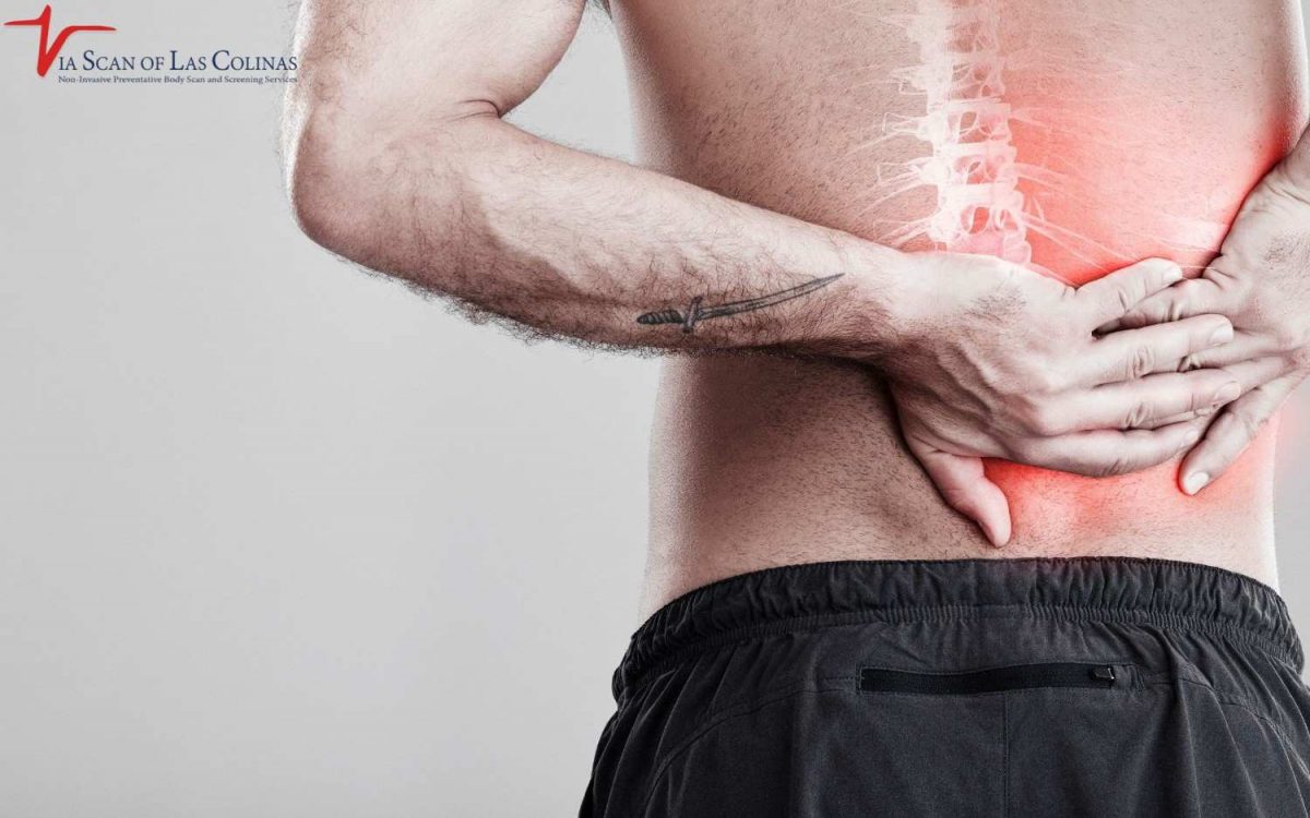

whole-body-scan-ViaScan-1200x750.jpg
02/Mar/2026

After a good night’s sleep, you wake up feeling pained in the back and ribs, and your day starts off frustrating. You are no longer refreshed and ready; you feel stiff, sore, and wonder why you wake up with a sore body after hours of sleep. Rib pain in the morning or rib cage pain after sleep is a common symptom in many individuals, and knowing the causes will assist you in concluding whether it is a mere sleep-related or a medical problem. Although it is normal to have temporary muscle stiffness after sleep, pain that is persistent or increasing is an indication of underlying conditions that require professional care. 

Why do I experience pain in my back and ribs upon waking up in the morning?

Back and rib pain in the mornings occurs because of different causes, including the mere problems with sleeping positions and some more severe medical disorders. Mattress support is poor, thus causing your spine to bow down in an unnatural manner resulting in pressure points to cause pain in the rib cage after you wake up. Natural issues such as arthritis, muscle injuries, or back complications tend to be more painful in the morning after rest. Whether it is normal to find the rib sore after waking up is a question that would depend on frequency and intensity, infrequent and mild pain is a common feature, but when pain is frequent and severe, it would be appropriate to seek the services of a professional.

Daily Habits That Lead to Overnight Pain

  • Sedentary life: Sitting in the same place hours results in stiffening of both muscles and joints and results in temporary pain in the ribs at mornings and back pain which often subsides with movement during the day.
  • Imperfect positioning of sleep: Sleep posture that twists the spine, intercostal muscle strain, or joint misalignment puts pressure on the thoracic cage, which accumulates when one sleeps, leading to pain on awakening which can be found till the muscles relax and restart positioning.

When Mornings Stiffness Is a Red Flag.

  • Constant intense pain: Morning pain that does not dissipate in the first 30 minutes of waking, or gets worse throughout the weeks or disrupts daily living might be a sign of chronic back pain causes that need medical imaging test.
  • Companion symptoms: Pain with fever, unexplainable weight loss, shortness of breath, or weakness is an indication of severe disorders that need immediate medical evaluation involving diagnostic tests such as CT scans.

Am I experiencing back and rib pain because of my sleeping position?

Of course, one of the most frequent causes of rib cage pains after sleeping and morning back discomfort is the sleeping position. Stomach sleeping causes your neck to bend sideways hours, the muscles of the neck and upper back work hard and the rib cage is positioned at an awkward angle. Sleeping on the side without proper support enables the top shoulder to roll forward, squeezing the rib cage and forming an imbalance in the body structure.

Even the back sleeping position will be troublesome in case your pillow is either too high or too low, thus forcing your back into unnatural positions. Being aware of what positions cause strain will enable you to make the necessary adjustments that could decrease the level of muscle tightness following sleep and prevents the occurrence of rib pains in the mornings before it occurs.

Is my mattress/pillow causing any back and rib pain in the morning?

Surely, that is true- your mattress and pillow make a huge difference in you waking up pain-free or achy. A mattress that is way too soft will result in an uneven sinking of your body, which results in misalignment of your spine and pressure points that will lead to you feeling some pain after sleep in your rib cage. A mattress that is too firm does not provide adequate cushioning of pressure areas, causing pain in certain body parts such as shoulders and hips and extending the pain to other parts such as ribs and back. The old mattresses become soft and form sagging areas which make your back bend in unhealthy postures when sleeping. Likewise, the pillows which are not suitable to your sleeping position do not provide the neck and spine with the right position during the night.

The selection of the Pillow and Firmness.

  • Back sleepers: require medium-firm mattresses with medium pillow height to maintain the natural curve of the neck without either pushing the head too far forward or letting it fall back.
  • Side sleepers: prefer medium to medium-hard mattresses with firmer and higher pillows to fill the space between shoulder and head to maintain the spine straight as opposed to curved.
  • Stomach sleepers: A firm mattress that is either very thin or does not have pillows should be used, and this position will cause the least amount of neck strain but this position causes the most trouble and should be avoided when possible.

What are the medical causes of back and rib pain after sleeping?

There are a number of health issues which make the back and the ribs achy in the morning and do not go away with a change in position or the purchase of a better sleep appliance. Spinal arthritis or arthritis in the rib joints causes inflammation and stiffness which exacerbates it following intervals of immobility during sleep. The consequences of fibromyalgia are widespread pains and muscle rigidity following sleep across the body such as the back, and ribs. The strains or even tears in the muscles that occurred during the daily activities are usually more painful in the morning when the muscles tightened during the night. If these chronic symptoms persist despite improving your sleep environment, it may be time to following sleep improvement would be a reason to begin inquiring about the causes of chronic back pain by seeking professional medical attention and diagnostic tests where necessary.

Kidney, Lung or Spinal Conditions.

  • Lung issues: Conditions like pleurisy can cause sharp rib pain that intensifies when breathing or changing positions during sleep
  • Kidney issues: Back and rib pain is common and is usually more painful on one side which is then accompanied by urinary complications, Diagnostic imaging can help clinicians rule out internal causes like kidney stones or infections.
  • Spinal problems: Herniated disc, spinal stenosis, scoliosis, or vertebral fracture cause chronic back pain that extends to the ribs – whole-body x-rays and specific CT scans assist in determining such structural issues that need medical care.

Conclusion

Even when you have intractable morning back and rib pain that does not seem to diminish with changing your sleep position or your mattress, professional diagnostic imaging can be of help to determine the underlying causes. We are ViaScan located at Irving, TX and we offer high-quality whole-body scan and CT scan services, which help medical professionals to diagnose conditions, that cause chronic pain, correctly.


cyst-vs.-tumore-e1770622956380.jpg
09/Feb/2026

When you find out that your doctor has ordered a CT scan due to the doctor finding a mass or a problem in your body can be frightening and intimidating. You may quickly start thinking about the worst-case scenario, but here is an important point to remember: not all masses are dangerous. Learning to distinguish the difference between cyst and tumor results on imaging will make you feel more knowledgeable and less fearful about what your scan results will show. The way cysts and tumors present on the CT scans is different, and the difference in the presentation will guide the radiologists in what they are dealing with. While some masses may require monitoring or further assessment, others may be harmless cysts that do not require treatment.

What is the distinction between a cyst and a tumor on a CT scan?

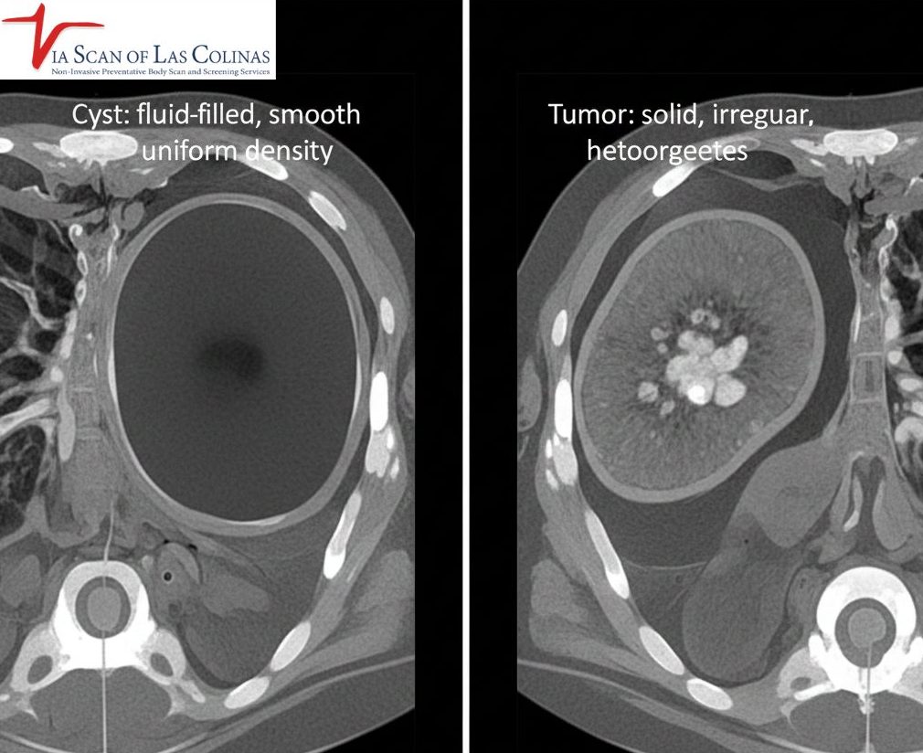
The very distinction between cyst and tumor on CT images is reduced to the contents of the mass. A cyst is a fluid-filled bubble – imagine it is a small water balloon in your body. In case of a CT, cysts are observed to be round and dark since they are filled with liquid, and not solid tissue. Tumors are abnormal growths of tissue that may be solid or contain both solid and fluid components. Tumors do not appear the same on CT scans they look different depending on the type of tumor, the tissue density and whether contrast is used. [1].

Can a CYST be Cancerous?

Most simple cysts are benign; however, some complex cysts with thick walls, internal partitions, or solid components may require further examination to assess whether they are potentially cancerous.

What would a cyst normally appear like on a CT Simple?

Simple cysts appear very characteristic on a CT scan and enable radiologists to identify them fast and be sure about them. Cysts are smooth, oval or rounded, and well delimited between the edges; they are clean and neat-looking and not irregular and jagged. Since they contain fluid, Cysts usually appear uniform in density on CT imaging, similar to fluid. Another characteristic that distinguishes them is that they do not enhance when contrast material is injected during scanning. Simple cysts have thin walls, so that they are barely visible, and the contents are homogenous, free of solid elements. These features may help radiologists identify a typical simple cyst, which is generally benign and does not usually require treatment.

Features That May Suggest a Simple Cyst 

  • Round form: Circular or oval edges that are not irregular.
  • Uniform fluid density: the same low (dark) density throughout.
  • Thin walls: Essentially, it is barely visible or very thin outer lining.
  • No enhancement: does not brighten when contrast dye is given.
  • No hard parts: There are no solid parts or pieces of any tissue present.
  • Borders: The cyst has clear edges, distinct borderlines between the cyst and the surrounding tissue.
  • Uniform internal appearance: Even internal structure throughout.

What are the ways of identifying and assessing tumors on a CT scan?

Tumors appear very dissimilar to simple cysts in CT scans and the dissimilarity helps the radiologists assess the type of mass they are viewing. The solid tumors are brighter or more heterogeneous when compared to the dark and uniform cysts filled with fluid. In the issue of cyst vs tumor identification, radiologists seek masses that are irregular in shape, lack smooth edges or finger-like projections protruding into the surrounding tissues- things that cysts do not possess. Lots of tumors improve tremendously when contrast material is injected that is, they become bright on the scan as the solid tissue will absorb the contrast. Some mixed masses are not entirely solid or liquid and need close consideration. 

Solid vs. Mixed Mass Features

  • Solid tumors: These masses are mainly composed of tissue rather than simple fluid. They can have different shapes, densities, and enhancement patterns on CT scans.
  • Mixed or complex masses: These masses contain both solid and fluid components and may appear partly dark and partly bright on CT scans. Depending on their features and location, additional imaging, follow-up, or biopsy may be recommended.

Does a CT scan effectively determine the benignity and malignancy of a mass?

CT scan provide exceptionally useful data on the masses but they cannot always be conclusive in diagnosing whether a tumor is cancer or not. In comparing cyst vs tumor results, CT imaging proves to be very successful in identifying simple and benign cysts that have clear fluid nature. In solid tumors, CT images reveal size, shape, location and relation to surrounding structures- all significant pointers to diagnosis. Some of the features involve increased risk: irregularity of borders, infiltration into surrounding tissues, quick growth between the scans, or unusual enhancement patterns. Yet, the question remains, is whether a cyst could be cancerous or whether it is a solid tumo and in this case, imaging alone may not always suffice. 

Limits of CT Imaging

  • Unable to tell the type of cell: CT is able to show the structure of the mass, but is unable to tell whether the cells are cancerous or not.
  • Similarity in appearance: There are benign and malignant tumors which have similar appearances on imaging.
  • Imaging findings should be correlated with the findings and medical history: Imaging findings must be correlated with symptoms and medical history.
  • Biopsy may be needed: Some masses can be characterized through imaging and follow-up, while a biopsy is considered when a definite diagnosis is needed. [1]
  • Very small abnormalities may be missed: CT scans have limitations in detecting very small lesions or microscopic changes.
  • Variability of enhancement: Enhancement can vary both in benign and malignant masses.
  • The follow-up can be required: Tracking scans over time can be used to assess growth patterns and behavior.

Conclusion

When your doctor has found out that there is a mass or an abnormality, the first step in the right direction to know what you are talking about is to have proper imaging. Your doctor will decide which scan is needed and whether contrast should be used that healthcare providers require to make a good differentiation between cysts, tumors, and other results. 

References:

[1] RadiologyInfo (Radiological Society of North America and American College of Radiology) — Abdominal and Pelvic CT. https://www.radiologyinfo.org/en/info/abdominct


dietery-instructions.jpg
05/Jan/2026

When you have a CT scan scheduled, you may be questioning yourself about whether you should have something to eat first. This is probably the most frequently asked question from patients,  and the answer varies according to what kind of scan you are having. Knowing how to prepare well will contribute to getting the results of your image as clear and accurate as possible. We would like our patients at ViaScan in Irving, TX to be certain and ready to attend their CT scan appointments. We will take a tour of the eating rules so you can have an idea of what to expect and how to get ready to have a successful imaging.

Can you eat before a CT scan?

The answer will depend on the kind of CT scan you are to undergo. There are CT scans that you can have a meal prior to the scan, and those that involve a fasting period of several hours. The instructions that you will receive will depend on the part of your body to be imaged and whether contrast material will be used or not. These guidelines are also worth following since they contribute to the generation of the clearest images. At ViaScan, we have professional staff that give preparation instructions as you are booking your appointment, so that as you reach our facility in Irving, you are well aware of what you need to do.

General Preparation Dietary Instructions Before CT Scan.

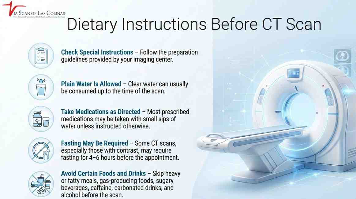
  • Check your special instructions: There are no special instructions that you are to follow, but always follow the instructions given to you by your imaging facility.
  • Plain water is generally permitted: The majority of CT scans allow you to take clear water until the time of the scan.
  • Timing of medication: The Majority of the prescribed medications can be taken normally with a few swallows of water.

Will consumption of food prior to a CT scan interfere with the accuracy of outcomes?

Yes, eating before some types of CT scans is known to influence the quality of your images. Food in your digestive system may complicate the imaging exercise of some organs. When food is in your stomach and in your intestines, it will look different on the scan compared to when they are empty. This variation may at times conceal valuable information that physicians should observe. To perform a CT scan in the presence of contrast medium having food inside your stomach can also affect the movement of the contrast medium inside your body. It is the reason why the medical community encourages the use of preparation guidelines to the letter because it directly affects the utility of your imaging findings.

Impact on Imaging Quality

  • Organ visibility: The presence of food may hinder the sight of such abdominal organs as the liver, pancreas, and kidneys.
  • Contrast distribution:  With an empty stomach, The contrast material can be better tolerated and imaging artifacts are reduced.
  • Image clarity; fasting decreases motion in the digestive tract, which makes images clearer and sharper.
  • Diagnostic Accuracy: Radiologists are more likely to detect abnormalities when the image is clear

Organs that are most susceptible to food consumption

  • Stomach and Intestine: These organs are literally filled with food and the appearance of the stomach and intestines alters because of the food.
  • Pancreas: It is an organ that is situated behind the stomach, and may be more difficult to look at when the stomach is full.
  • Gallbladder: Food activates digestive processes  and this can have a change in the appearance of this organ.
  • Blood vessels: Blood vessels in the abdomen can be more readily observed clearly when other organs are empty.

What are the differences in preparation rules in CT scans with contrast and without contrast?

There are two types of CT scans: CT with contrast material and CT without contrast material. These two types have vast differences in the preparation requirements. Knowing the kind of scan you are getting makes you ready accordingly. At ViaScan, we provide contrast and non-contrast CT scans, including full body scans, with well-written instructions that are specific to each scanning type, to the community of Irving, TX.

Non-Contrast CT Preparation

There are fewer dietary restrictions in non-contrast CT scans, with most non-contrast scans, particularly ones that involve your head, chest, or extremities that you can typically eat and drink prior to your CT scan procedure. Nevertheless, despite the non-contrast abdominal scans, other facilities might ask patients to undergo light fasting to increase the quality of the image. The most important difference is that in the absence of contrast material, the risk of nausea is ruled out, and the only issue is that the area of interest is clearly visualized. It is always important to ask your imaging center whether or not your particular non-contrast scan needs any dietary alterations.

CT Scan With Contrast: Fasting Guidelines.

  • Four to six-hour fast: The majority of CT scans with contrast would ask you to avoid food during this time before your appointment.
  • Clear liquids permitted: Water and perhaps clear broths may be allowed up to two hours prior to the scan.
  • Medication considerations: Take all applicable medications with water in little sips unless otherwise instructed.
  • Contrast safety: Fasting decreases the chances of nausea or vomiting during contrast.
  • Hydration value: It is very important to drink a lot of water before the fasting period commences so that your kidneys can properly filter and eliminate the contrast.

What do you avoid eating or drinking before a CT scan?

 As a rule, you are not supposed to eat heavy, fatty foods within the hours before your scan, even without fasting. These foods have slower rates of digestion and it may stay in your digestive system when you have imaging. Foods that produce gas, such as beans, carbonated drinks, and some vegetables, may also cause air pockets, which influence the image of the object being photographed. Our professional staff at ViaScan assists the residents of Irving in knowing which objects to avoid, given their respective scan needs.

Foods That Can Interfere with Imaging.

Foods that form gas in your digestive system, like broccoli, cabbage, and beans, may result in air bubbles that will show up on the scan and may conceal key anatomical features. People with sensitivities might sometimes have the same problem with dairy products. In case you are allowed to eat, just eat light and easily digested food such as toast, crackers, or plain rice. These basic foods are energy-giving without the development of imaging complications.

Beverage Restrictions

  • Stay away from sugary beverages: Soda and fruit juices may influence abdominal imaging.
  • Avoid Caffeine : Coffee or vitality can enhance digestive processes.
  • No Alcohol: You should abstain from alcohol at least 24 hours prior to your scan.
  • Carbonated drinks. These generate gas, which may interfere with images.
  • Water is the best: The safest is plain water, and it can be used to eliminate contrasts.

Conclusion

Correct CT imaging results require proper preparation. ViaScan is a laboratory where skilled radiologic technologists offer individualized preparation guidelines to all patients who would like to have CT scan services, including whole-body scan options, in Irving, TX. We will ensure the imaging experience is comfortable and stress-free, and make sure we provide the best diagnostic images.


CT-scan-showing-kidney-stones-monitored-ViaScan.jpg
29/Dec/2025

The pain caused by kidney stones can be severe and varies depending on the individual and the stone’s characteristics. The first question people will pose is: How long does it take for a kidney stone to pass? The response is relative, just like in the case of individuals, and it depends on a number of factors such as the size and the position of the stone. The knowledge of the influence of passage time will allow you to determine what to expect and when to consult professional imaging. For individuals who need imaging to assess kidney stones,advanced CT scans can accurately identify and track the kidney stones’ size and location along the urinary tract.

What is the average time for a kidney stone to pass?

There is no guaranteed timeline. In general, smaller stones are more likely to pass on their own and tend to pass sooner, while larger stones are less likely to pass without treatment. [2] Some small stones pass within days, while others take weeks, and some never pass without help. Because passage time is so unpredictable, medical evaluation and follow-up are important. Your doctor can confirm the stone’s size and location, check whether it is blocking the flow of urine, and advise whether waiting for it to pass is safe or whether treatment is needed.

What Determines How Quickly a Kidney Stone Passes

  • Stone size- smaller stones will move faster than bigger stones.
  • Presence of a stone in the urinary tract.
  • Anatomical and size differences in the ureters of individuals.
  • Fluid and hydration status.
  • Movement patterns and physical activity.
  • Stone composition (e.g., calcium oxalate, uric acid)
  • Past history of kidney stones.
  • Urinary tract constriction or obstruction.

What is the impact of the size and location of a kidney stone on the passage time?

Size and location are the two biggest factors in whether a stone will pass on its own, but they can’t predict an exact timeline. [2] Kidney stone size plays an important role in how quickly a stone may pass. Smaller stones are more likely to pass naturally, while larger stones may take longer or require medical treatment. Although some stones pass within a few days, others may take several weeks. The exact time depends on the stone’s size, location, and whether it is blocking the urinary tract.  Place is also important,–stones in the kidney take a longer time to reach the bladder than those already in the ureter. 

What are the signs that a kidney stone is moving or is going to pass?

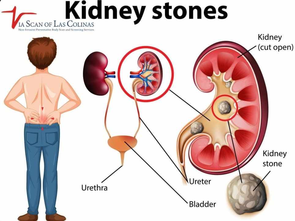
Pain that starts in the back or side and then moves toward the lower belly or groin is common as a stone travels down the ureter. [1] .

The urgency and difficulty to urinate, which is then followed by real urination, could be a sign of the stone approaching the bladder. Temporary relief often occurs once the stone enters the bladder, as the bladder is more distensible than the narrow ureter. But with fresh pain, we can get where the stone comes out of the bladder and through the urethra. The knowledge of these patterns will make you aware of when you are making progress on the passage. 

Common Warning Signs

  • Acute pain, which radiates in the back or side and into the lower abdomen and groin.
  • Urgent and frequent urination.
  • Painful urination.
  • Urine of pink, red, or brown color with evidence of blood.
  • Blurred or smelly urine.
  • Pain changes with associated vomiting or nausea.
  • Fever or chills, which can mean an infection: get medical care the same day, because an infected, blocked kidney is an emergency.

When Pain Patterns Change

Suddenly intensified pain can indicate that the stone is lodging in a small section of the ureter. Pain that shifts toward the lower abdomen or groin may occur as a kidney stone moves through the urinary tract. However, changes in pain location do not confirm that the stone has reached the bladder.

A sudden, sharp pain during urination may indicate the stone is passing through the urethra, often the final stage before complete expulsion. Nevertheless, others feel very little pain during the process, and others end up feeling very uncomfortable for weeks. 

Is there a way to accelerate the natural passage of a kidney stone?

There is no way to make a stone pass on a set schedule. For small stones, your doctor may advise drinking enough fluids to keep your urine pale, but drinking very large amounts of water will not force a stone past a blockage. [2] You cannot make a kidney stone pass on some timeline, but there are a few measures that can be taken to help smaller stones pass on their own. Drinking enough water to stay hydrated may support the natural passage of small kidney stones. However, drinking excessive amounts of water will not necessarily make a stone pass faster. Gentle activities, such as walking, may be comfortable for some people, but they do not guarantee faster stone passage. The methods can only succeed with small stones, however, which are already likely to be passed naturally. The duration of kidney stone passage using these methods is still highly varied. 

Hydration and Movement Tips

  • Drink water regularly during the day, no longer only when you are thirsty.
  • Goal: clear or very pale yellow urine as an indicator of hydration.
  • Avoid caffeine and alcohol, which can cause dehydration.
  • Keep track of the fluid consumption to guarantee a sufficient level of hydration.

Dietary Options

Other medical practitioners prescribe citrus juices and citrus fruits containing citrate that can be used to prevent the formation of stones. Restricted sodium intake is beneficial in lowering calcium in urine. There is the possibility of avoiding high amounts of oxalates in foods such as spinach, chocolate, and nuts based on the type of stone. But the food will not help a lot to increase the speed of passage of the old stones–they are more preventive. Professional consultation will make sure that dietary advice is in keeping with your type of stone and health condition.

Conclusion

The benefit of knowing the schedules of passing kidney stones is to relieve expectations in an unpleasant situation. Although small stones may just pass out of their own accord within days to weeks, proper detection and observation will help you determine whether natural passage is on the right track or medical treatment is to be adopted. The scanning services offered at ViaScan under the supervision of John Duncan include the full body wellness scan and CT scan, which give a close-up image of the location of your stone.

Resources:

[1] National Institute of Diabetes and Digestive and Kidney Diseases (NIDDK) — Symptoms & Causes of Kidney Stones. https://www.niddk.nih.gov/health-information/urologic-diseases/kidney-stones/symptoms-causes

[2] National Institute of Diabetes and Digestive and Kidney Diseases (NIDDK) — Treatment for Kidney Stones. https://www.niddk.nih.gov/health-information/urologic-diseases/kidney-stones/treatment


CT-Scan.webp
22/Sep/2025

Medical imaging has advanced well beyond mere X-rays, and advanced machines are now available that can ideally view the inside of the body. One of the most beneficial tools for physicians today is the CT scan, which may be performed with contrast when additional detail is needed. Contrast-enhanced CT can help clinicians assess certain organs, blood vessels and tissues. The scan itself is usually quick, but its suitability and risks depend on the patient and the type of examination.

Contrast can make selected structures easier to distinguish than on scans without contrast. ViaScan of Las Colinas provides several imaging services; however, not every scan uses contrast material. We will discuss the special qualities of contrast CT scans and when they may be prescribed to you.

What is a CT scan with contrast, and how is it different from a traditional CT scan?

  • Normal CT images are clear in showing bones and certain organs. 
  • Contrast material works like a highlighter. It does not permanently colour your organs, but it temporarily changes how X-rays pass through blood vessels, organs and tissues, so they stand out more clearly on the images. [1].
  • The contrast agent travels through your bloodstream, which helps blood vessels and certain organs and tissues show up more clearly.
  • A contrast-enhanced CT can give the doctor extra detail for some questions, but it is not automatically better for every purpose.

What is contrast dye made of, and how does it work?

Iodine-based contrast material is commonly used for CT scans. It functions as a highlighter for your insides. Once injected, the dye spreads in the blood and results in certain tissues appearing brighter in CT images. Iodine absorbs X-rays in a different way to increase the contrast of structures. The dye is filtered out by your kidneys and passed in urine; how quickly this happens varies from person to person and may take longer if kidney function is reduced. The imaging team follows procedures for giving contrast and responding to any side effects or reactions.

When do doctors recommend a CT scan with contrast?

When physicians need to see blood vessels, detect infections, or examine soft tissues more clearly, they may prescribe contrast. A CT scan without contrast already shows bones and some organs well, but contrast helps doctors see problems in soft tissues and blood vessels that would otherwise be difficult to detect.

If your doctor suspects a problem with blood circulation, is looking for tumours, or simply wants to examine the inside of your kidneys, liver, or other organs, they may request a contrast scan. Contrast is often used for detailed scans of the blood vessels, heart, and lungs, but many preventive scans, such as a coronary calcium score, are done without any contrast at all.

What conditions can a CT scan with contrast help detect more clearly?

A CT scan enhanced with contrast can help doctors see certain health issues more clearly than a standard scan.

It can help doctors:

  • investigate blood clots, when the right type of CT exam is used,
  • assess blood vessels,
  • look for signs of infection. CT shows the structure of organs; it does not directly measure how well they function.

Contrast can help doctors identify obstructions in your blood vessels or veins, as well as issues with your kidneys, liver, or how blood circulates to various body parts. Contrast scans are usually ordered by a doctor to investigate a specific problem. Separately, ViaScan offers preventive wellness scans for people without symptoms, and your doctor can help decide whether a contrast scan is ever needed as part of your care.

Is a CT scan with contrast safe?

A contrast-enhanced CT scan procedure is safely performed on most people, but your doctor will review your medical history first. Before your scan, tell your doctor if you have had a reaction to contrast before, have allergies to food, drugs, or other substances, or have heart disease, diabetes, kidney disease, or thyroid problems, because these can affect how your scan is planned. [1] 

If you are or might be pregnant, always tell your doctor or technologist first; iodine-based CT contrast is not known to pose a significant risk to mother or baby, but your radiologist can explain the risks and benefits. [1].

Where can I get my health scanning done?

At ViaScan, we do not just take pictures. We provide imaging services, including heart scan, lung scan, and wellness body scans. Our standard heart and body scans do not use contrast dye, so contact us to confirm whether a specific contrast-enhanced CT is available. Scanning can help patients stay informed about their health, with follow-up from a qualified healthcare professional.

Conclusion

Knowing what a contrast-enhanced CT scan involves can remove some of the mystery and anxiety that are sometimes associated with medical practice. Such scans are an effective tool that enables the doctor to have an amazing view of the inside of your body with high clarity and detail. The contrast enhancement introduces important data for proper diagnosis and treatment planning.

The power is in knowledge as far as your health is concerned. As an active participant in your healthcare choices, you learn about procedures such as contrast-enhanced CT scans. The professional staff of ViaScan of Las Colinas is ready to clarify any questions and help you feel at ease throughout the process.

References:

[1] RadiologyInfo (Radiological Society of North America and American College of Radiology) — Contrast Materials.https://www.radiologyinfo.org/en/info/safety-contrast


How-does-a-CT-scan-work.jpeg
11/Aug/2025

When a healthcare provider suggests a CT scan for diagnostic purposes, you might feel curious about it. It is completely common to have certain questions, such as what happens during a CT scan, whether it is risky, or whether it has any side effects. Knowing about the imaging process and how it works will make you feel more guided.

Let’s walk you through each step of the CT scan, from how it works to scan preparation to what happens during or after the procedure.  

What is a CT scan?

A CT scan is a medical imaging test that uses X-rays and computer processing to create cross-sectional images of the body. These images can also be combined into 3D views when doctors need them, but not every CT scan produces a 3D image. The images show bones, organs, soft tissues and blood vessels in much more detail than a standard X-ray. CT images are used to help diagnose injuries and diseases, including some cancers and infections, and to check how well a treatment is working.

How does CT imaging technology work?

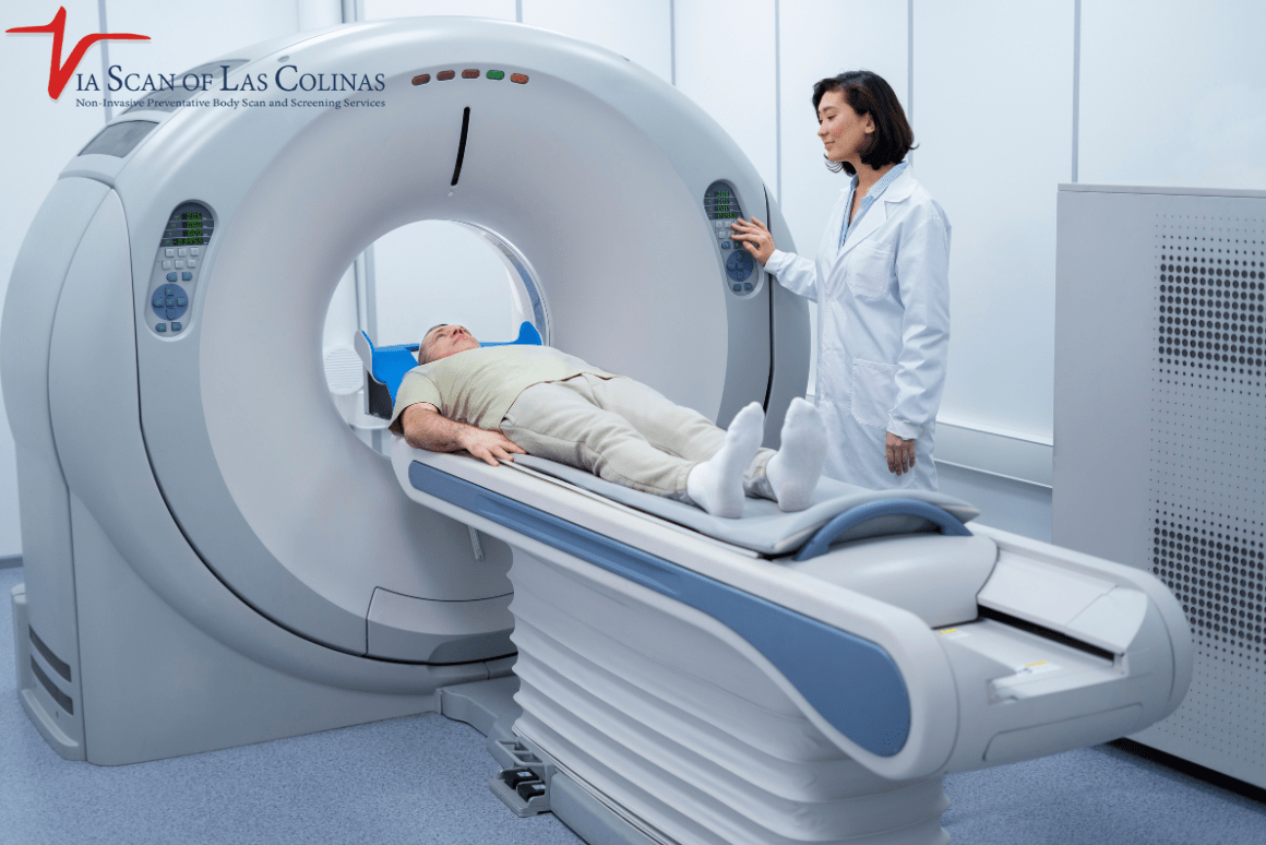
The CT scanner is a large, doughnut-shaped machine with a moving bed attached to it that will go in and out while taking pictures. 

X-ray source 

While you are lying down on the table, the X-ray source will move around your body, sending radiation to your targeted body parts.

Detectors

Advanced detectors will detect the emitted radiation on the other side of the body. 

Computer processing 

The computer will collect the data and combine it to create a 3D image that your healthcare provider can see on the screen. You can see it later as well. 

CT can show fine detail, such as small fractures and some small tumours, although no imaging test detects every abnormality.

Why do I need a CT scan?

Your doctor might suggest a CT scan for various reasons, including:

  • Detect bone fractures 
  • Identify any early tumours or blood clots 
  • Detect blockages in blood vessels 
  • Help evaluate suspected infections and certain heart conditions, although CT is not the right test for every type of heart disease 
  • Monitor the progress of treatment 
  • Trace disease progression 

Doctors often use CT when they need detailed images quickly.

How do I prepare for the CT scan?

The scan preparation depends on the scan your doctors have suggested for you. Moreover, your doctor will explain everything you need to know, from how you need to prepare for the scan to what will happen during the procedure. Here are some guidelines:

  • You may be advised to stop eating or drinking a few hours before the scan. 
  • Tell the imaging team if you have had a reaction to contrast before, or if you have allergies to medicines, food or other substances. [2].
  • Also tell them if you are or might be pregnant, have kidney disease or diabetes, or take medicines such as metformin. [2]
  • You must arrive early on the scan day to become familiar with the surroundings and remain calm. 
  • Remove all the jewellery before the scan, as any metallic object can distort the images, leading to inaccurate results.
  • It’s better to change into a hospital gown for a more comfortable journey.

Furthermore, if you have any questions or concerns, you can ask your doctor right away.

What should I expect during my CT scan?

  1. First, you will arrive at the facility, and your doctor will review all the instructions and re-explain the entire process to ensure there is no confusion.
  2. Some CT scans use a contrast agent, given by injection into a vein or as a drink, to make certain structures easier to see. Many scans, including coronary calcium scoring and low-dose lung screening, do not use contrast. You will be told in advance if your scan needs it.
  3. You will be asked to lie on the bed.
  4.  The bed will go inside the scanner during the scan.
  5. A CT technologist operates the scanner from an adjoining room and can see, hear, and speak with you throughout the scan.
  6. When the bed begins to move inside the scanner, you will hear some whirring sound as well.
  7. During the scanning, you may be asked to stop breathing. This will be only for a few seconds.

The computer will collect all the images, and your doctor can now see them for diagnostic purposes. A radiologist interprets the images and writes a report. Your own doctor then discusses the results and any next steps with you

How long does a CT scan take?

The scan itself is usually quick, but how long your whole visit takes depends on the type of scan and the preparation needed. For example, scans that need contrast, especially an oral contrast drink, can take longer. Ask your imaging facility how much time to plan for.

Are there any risks of a CT scan?

CT uses X-ray radiation. For any one person, the added cancer risk from a scan is small compared with the natural risk of cancer, but it is not zero, which is why a scan should be done only when the expected benefit outweighs the risk.[1]. However, if you have certain health concerns, you can talk to trained professionals for a clearer and better understanding. 

People often ask, “Can I get a CT scan while pregnant? Or does it have a side effect?” Scans without contrast usually cause no side effects. Contrast agents are not dyes; they are substances that temporarily change how X-rays pass through the body. [2] Precautions for people with kidney problems depend on how well their kidneys work and the type of contrast used. [2]

Conclusion 

A CT scan is more than just a diagnostic test; it provides clear answers and guides you to the right treatment options. It’s normal to have questions, and finding a trusted partner for a CT scan you can count on for your health is essential. At ViaScan, we offer low-dose CT scans and prioritise patient safety and comfort to ensure you receive the best care. Our trained professionals will answer all your queries and will walk you through each step.

If your doctor has recommended a CT scan, trust ViaScan under supervison of Dr. John Duncan offers CT imaging, including low-dose CT where appropriate, and our team can answer your questions about preparation and what to expect.

References:

[1] U.S. Food and Drug Administration — What are the Radiation Risks from CT? https://www.fda.gov/radiation-emitting-products/medical-x-ray-imaging/what-are-radiation-risks-ct

[2] RadiologyInfo (Radiological Society of North America and American College of Radiology) — Contrast. https://www.radiologyinfo.org/en/info/safety-contrast


heart-CT-scan.webp
21/Jul/2025

Time is of the essence when you have a heart to worry about! When you consult your doctor and are scheduled to get a Heart CT scan, you are likely to ask yourself how many hours of your day will be consumed. You said you are going to miss lunch? Can you fit it into your lunch hour? Are you free to clear your afternoon?

Being a qualified scanning expert, I receive these time-related questions daily. Customers prefer to know clearly what they will be signing up for, and frankly, that is good planning! A heart scan may appear to you to be something never-ending; however, the truth of the matter is more thrilling than what you can imagine.

The heart scanning of today is fast, convenient, and designed to suit your hectic lifestyle with minimal interruptions. Let us break down what you can really expect!

What is the Time Taken for The Heart CT Scan?

And here is the most amazing moment: most people would be surprised to learn that the actual heart CT scan itself can take only 10 to 15 minutes. 

Throughout those several seconds, you will lie on a comfortable table with everything still as the CT scanner will rotate about you and take hundreds of detailed images of your heart. The machine is fast enough to capture clear pictures between heartbeats, which is quite amazing when one takes a second to consider this.

In some cases, when your heartbeats are quick or irregular, it may take 20 minutes to scan, which is incredibly fast. The scanner has become so advanced that, intelligently, it can operate in sync with your heartbeat, allowing it to capture perfect pictures once.

The point is to remain quite stationary in those seconds. Consider it a very significant photograph; you want to be as still as possible so that the image turns out to be as clear as possible.

How long should a customer’s overall visit last?

Although the actual scanning itself is extremely rapid, you will spend between 30 minutes and an hour at the facility. This consists of the total time of preparation, which is equally important as the actual scan.

Upon arrival, you will need to complete some paperwork and allow 10 to 15 minutes for check-in. Next, you will be transferred into a comfortable hospital gown, a process that will take approximately five minutes. The technologist will discuss the procedure and address any questions you may have about it.

If you require the use of contrast dye, you may need an additional 10 to 15 minutes to prepare. The staff will also place certain monitoring electrodes on your chest to monitor your heart rhythm during the scan.

Once you have completed your “heart scan,” you will take a couple of minutes to dress and then be given basic instructions regarding your results. Most of the facilities are highly efficient, and your time is respected; therefore, they maintain a steady pace.

My advice? Allow for an hour in total and carry a good book or download a podcast in case of a minor delay.

Is the heart scan time-consuming with Contrast Dye?

Great question! Taking contrast dye does take some time from your appointment, but not if you believe. The contrast dye helps produce ultra-clear and detailed images of your heart and blood vessels, much like high-definition television does for regular tv.

An additional 15-20 minutes should be allowed when contrast dye is required. This will consider the time needed for a technologist to insert a small line into your arm using an IV, a procedure that lasts approximately one to two minutes and involves a slight pinch.

The contrast injection occurs immediately prior to your scan and usually requires no more than 30 seconds. You may experience a warm feeling that runs through your body – it is perfectly normal and, in fact, an indication that the contrast is working just as it should.

Once the scan is complete, you will lie down for approximately 10 minutes as the medical staff ensures you are in a stable condition before you leave. There is concern among some people about side effects; however, serious reactions are extremely rare with newer contrast materials.

The fortunate part is that the actual heart CT scan is no longer necessary due to the use of contrast; the 10 to 15 seconds it takes to scan the body part is still extremely fast.

Do I Immediately Get the Results After the Scan?

This is the most frequently asked question, and I completely understand the reasons why you would want to have the answers at hand. Although the scanning process is incredibly fast, it simply takes a little longer to get your total results, as precision is crucial when it comes to your heart.

All the pictures from your so-called heart scan will also be thoroughly studied by a specialised radiologist (read: a physician highly qualified in interpreting medical images). The elaborate review would typically require 24-48 hours, as they would scan hundreds of pictures to ensure nothing is overlooked.

Yet, in case something requires attention, the radiologist will call your doctor immediately, e.g. even when you are still in the scanning centre. The emergency findings are dealt with urgently; therefore, you never think of delays when something important is at stake.

Depending on the specifics of your doctor’s request, especially in certain parts of scanning centres, the preliminary results can be provided on the same day. The findings will then be discussed with you by your doctor, who will clarify them regarding your health status.

You will receive a follow-up in most places within a few days, or they may have an online patient portal where results are available.

What can I expect in Recovery?

Yet another piece of good news is that there is practically no recovery period or time required after a heart CT scan. Because it is a non-invasive process (that is, nothing is put inside your body, except that the milk chocolate and marble chocolate might remain outside of your body by dint of your IV and your stomach), you are able to resume all your usual activities immediately.

If you received contrast dye, you may be advised to drink additional water throughout the day to aid the absorption process, but this is the most specific advice that most people receive.

You can drive home, return to work, exercise, eat normally, and do what you usually do. One can perform any activity without limitation following a heart CT scan.

Others are slightly nervous after waiting to get their results, and this is normal. It is essential to remember that no news is typically good news; however, if you have any updates to share, please contact your medical team immediately.

Conclusion

Receiving a heart CT scan should not be the event that affects your whole day or makes you stressed over time. When it takes only 10 to 15 seconds of real scanning time and the total time is approximately an hour to gather a wealth of information regarding your heart’s health, it is among the most effective time-saving methods to find out in detail about your heart health.

At ViaScan, we appreciate your busy life, and we will serve you well through efficiency and quality in our heart scanning services. When it comes to protecting your heart, you cannot afford to lose any moment (and that’s why we make sure these moments are worth spending)


What-is-the-most-Common-reason-for-a-CT-scan.jpg
16/Jun/2025

There is no single most common reason for a CT scan. Doctors order CT scans for many different reasons, most often to investigate symptoms such as abdominal or chest pain, to check for injuries after an accident, to look for problems in the head or brain, and to help diagnose and monitor conditions such as infections and cancer. [1]

CT scans may be something that one can easily think of as terrifying; however, when creating this impression, one is referring to one of the most useful tools available to doctors today. Imagine them as something like super-powered cameras that can look inside your body, and they do not even need to cut you open. We’re going to dive in and look at everything you want to know about these amazing medical images!

What is a CT scan? Why Is It so Common?

Computed Tomography, or just CT. Imagine it as a special X-ray, but with multiple pictures taken from various angles. Consider, for example, that you would like to find out what is inside a gift that has been wrapped in paper but has not been opened yet. An X-ray is akin to shining a flashlight at a specific angle – you can still see some shadows, but the entire image will not be visible. A CT scan is the equivalent of dozens of flashlights being shone in all directions, allowing a person to see inside and out of everything.

Why Are CT Scans So Popular?

They’re Fast

The average procedure on most CT scans requires only 10-30 minutes. It’s not as much time as you’d need to watch your favourite TV show!

They’re Detailed

CT scans can detect what is not visible even in regular X-rays. They are able to see the problems hiding behind the bones, or in other places which are not easily visible.

They do not hurt

CT scans are painless, although they do use X-ray radiation (see below). [1]  All you need to do is lie on a table and let the machine do its job.

They Assist Doctors to Make Rush Decisions

A CT scan can give doctors a quick insight into what is wrong with the patient, especially when such a patient visits the emergency room with critical symptoms.

What Is the Most Common Reason to Get a CT Scan?

CT scans explore organs such as your liver, kidneys, pancreas, and intestines. What is the reason behind the prevalence of pelvic scanning? CT scans of the abdomen and pelvis are often used to find the cause of belly or pelvic pain and to check organs such as the liver, kidneys, pancreas and intestines for infection, inflammation, injury or other problems. Because many organs sit close together in the abdomen, imaging can help doctors pinpoint which one is involved.

Why Does a Doctor Order a CT Scan?

Your doctor decides whether a CT scan is needed based on your symptoms, examination and medical history. Having one of the symptoms below does not automatically mean you need a scan.

Brain Scans and Head Scans

These are overly common, as well! The brain CT scans are ordered by a doctor when individuals experience the following:

  • Unrelenting bad headache
  • Light-headedness or dizziness, Dizziness or balance problems
  • Memory issues
  • Accident or fall, head injury

Chest Scans

The place that houses your heart and lungs is your chest. Physicians may request a chest CT scan as a measure to check:

  • Breathing problems
  • Chest pain
  • Non-productive coughing
  • Lung infections

The screening for cancer

CT scans are scavengers in the hunt for cancer cells. They use the ability to assist doctors:

  • Identify the tumours that may be hiding
  • Check whether the therapy against cancer is effective
  • Ensure whether it has spread to other parts of the body due to cancer.

Taking a glance at Injuries

CT scans can be used to quickly demonstrate when you are involved in an accident or if you hurt yourself:

  • Broken bones
  • Internal bleeding
  • Organs destruction
  • Blood vessel issues

Wellness and Prevention

The number of people undergoing CT scans, even when they do not feel unwell, is increasing. The FDA notes that whole-body CT screening of people without symptoms has not been shown to meet accepted criteria for an effective screening test, and that findings can lead to further tests. Some targeted screening scans, such as low-dose CT lung screening for eligible adults, are recommended by guidelines. Discuss any preventive scan with your doctor. [2][3]

What Do I Need to Do to Prepare for My CT Scan?

Preparation of a “CT scan” is generally quite simple! This is what you must know:

Check Your Rules of Eating

In some CT scans, you will need to avoid eating for several hours prior to the scan. Your physician will inform you about the necessary steps, such as fasting before a blood test – not an exciting prospect, but not difficult, either.

Take plenty of water.

Drink plenty of water, unless your doctor advises otherwise. This helps the kidneys function more efficiently and, in some cases, makes the scan clearer.

The Day of Your Scans

Do not Wear Tight Clothes

Opt for clothes that lack metallic zips, buttons, and undergarments. Metal appears in CT pictures and may prevent the view. Plump into pyjamas or sweatpants!

Take out Metals

Remove jewellery, watches, and all other pieces of metal. Even minor items such as bobby pins will influence the pictures.

Carry Your Medical Data

Carry a list of your medications and scan results from the past. It would be comparable to a person showing up to school with their homework, a positive way of helping others complete their work more efficiently.

Contrast Dye

In some cases, doctors may want to apply a special type of dye to enhance the appearance of a specific portion of your body in the scan. And don’t worry – it is normal.

Drinking Contrast

Before you have a scan, you may be required to take a chalky drink. It doesn’t have a particularly good taste, but neither is it particularly bad. Just imagine it’s a strange kind of milkshake that helps your doctor see better.

IV Contrast

In other cases, they will inject the dye into your arm using a small needle. You may experience a warm feeling or a strange taste in your mouth for several minutes. There is nothing wrong with this!

Is the CT Safe? What Risks are involved?

This is likely the question that causes the greatest concern for most people, and it is entirely normal to feel anxious about it. What is the situation? “CT scans” are safe. Each year, millions of people acquire them without difficulties. There are virtually always more pros than minor cons associated with learning about the medical problems ailing your body.

Radiation

When you are flying, you will be exposed to a small amount of space radiation. A CT scan delivers more radiation than a plain X-ray; the amount depends on the type of scan, and low-dose scans use less. [1]

The Risk Is Very Minor

For any one person, the added cancer risk from a CT scan is small compared with the natural risk of cancer. The FDA estimates that a scan with a dose of 10 mSv may raise a person’s risk of fatal cancer in the US is about 400 in 2,000 to about 401 in 2,000. That is why CT should be used when the benefit outweighs the risk. [1]

What do the Results Mean?

Here is what your results may mean: This could be what could befall it:

Normal Results

When analysis reveals that everything appears to be normal, your doctor will inform you that the scan will not reveal anything. That is good news!

Abnormal Results

In the case that something abnormal is detected in the scan, do not freak out! Not everything labelled as abnormal is inherently bad. It simply may imply:

  • It is just a small infection that can be cured easily
  • It is something which must be monitored over time
  • You will require another test to have a better picture

Follow-Up Tests

Your doctor may occasionally need to conduct additional tests to gather more information. This resembles taking a second opinion – it is used to ensure that they get the full picture.

Conclusion 

Whether you are considering a wellness CT scan or your physician has prescribed it, ViaScan under the supervision of John Duncan, Ph.D., offers a variety of non-invasive preventative body scans and screening services, ready to make the CT scan process as easy and informative as possible. We understand that undergoing any medical test can be intimidating, but it doesn’t have to be. Whether you are undergoing a scan at your doctor’s request or not, we will guide you through all the procedures. We are eager to address all your inquiries and clarify everything in simple, easy-to-understand language.

References:

[1] U.S. Food and Drug Administration — What are the Radiation Risks from CT? https://www.fda.gov/radiation-emitting-products/medical-x-ray-imaging/what-are-radiation-risks-ct

[2] U.S. Food and Drug Administration — Full-Body CT Scans: What You Need to Know. https://www.fda.gov/radiation-emitting-products/medical-x-ray-imaging/full-body-ct-scans-what-you-need-know

[3] U.S. Preventive Services Task Force — Lung Cancer: Screening (Final Recommendation, 2021). https://www.uspreventiveservicestaskforce.org/uspstf/recommendation/lung-cancer-screening


Difference-between-CT-scan-and-MRI-1.jpg
11/Jun/2025

You are in the doctor’s facility, and they say that you require a scan. Suddenly, your mind begins to race with questions. CT scan? MRI? What is the distinction? Will that be painful? Should I fast?  Don’t worry! We are going to make you an expert in scanning without giving you a headache. Just imagine this is your introduction to these wonderful machines that look inside your body like X-ray vision superheroes.

Why are there so many Scans, and what is so Popular about CT and MRI?

Your body is a complicated puzzle with numerous pieces. There are cases where doctors require additional equipment to visualise other parts.You can imagine it like when you take photos. There are times when you use your phone camera to take impromptu shots. Then there are times when you require a complex camera with specific lenses to capture close-up images. The CT scans and the MRI machines are like those various cameras – each of them is truly excellent at demonstrating things inside your body.

The most popular are CT scans and MRIs, as they are the Swiss Army knives of medical imaging. They are able to visualise many different things and assist the doctors to know what is going on inside you without necessarily having to operate on you first.

What is the CT Scan vs. MRI Difference?

The key difference: CT scans use X-rays, while MRI scans use a strong magnet and radio waves. Unlike CT scans, MRI does not use ionizing radiation. [2]

CT Scan:

  • Involves X-rays that revolve around you.
  • Composite photographs in slices, as if they were slicing bread
  • Super quick – often takes only a few minutes
  • You are put on a table which moves inside a large doughnut-shaped machine

MRI:

  • The category utilises strong magnets and radio frequencies.
  • Slices pictures as well, with better soft-tissue contrast
  • Time-consuming – generally takes longer, depending on the examination
  • Produces thunderous knocking and buzzing 
  • You are lying in a tube-shaped machine.
  • They are both fantastic at their work, simply in different ways.

Which is safer, a CT Scan or an MRI?

This is what worries you the most. Both CT and MRI scans are generally safe when medically appropriate, but they involve different risks and precautions.

CT scans use ionizing radiation. A typical chest CT, for example, gives about the same radiation dose as two years of natural background radiation. [1] Doctors use the lowest radiation dose needed to obtain useful images and avoid unnecessary repeat scans.

MRI scans do not use ionizing radiation. Radio waves and magnets are generally safe for most individuals. Some implants and devices, such as certain pacemakers, may not be safe in the MRI environment, while others are. Always tell the imaging staff about any implant or medical device before the scan. [2]

Your doctor can explain why a scan is recommended, what benefits it offers, and whether it is the right test for your condition.

What is Best Diagnosed with Which Type?

This is the place where these machines can really demonstrate their superpowers!

CT scans are excellent when:

  • Fractures and injuries
  • Trouble in your chest, belly and pelvis
  • Finding infections
  • Fast examination of your organs
  • Emergencies where answers are required quickly by doctors

MRI scans excel at:

  • Your brain and your spine
  • Examining muscles, tendons and ligaments
  • The visualisation of soft tissues at close-up view
  • Locating issues in the knees and shoulder joints
  • Obtaining truly clear images when physicians must view small details

Most of us have questions like, Can a CT scan detect cancer? And the answer is yes. CT scans can help detect and evaluate many types of cancer. They can show tumors, help determine whether cancer has spread, and assist doctors in planning treatment. MRI can also help detect and evaluate certain cancers, particularly in areas such as the brain, spine, and soft tissues.

What About Contrast Dye – Do the Two Tests Use It?

Now and then, yes; now and then, no! Contrast dye is like having special paint in your body that helps some areas show up better in the pictures.

If you are undergoing a CT scan, you may be given a liquid contrast solution to drink or receive an injection into a vein. There’s nothing to be afraid of – according to most people, it only makes them feel warm for a few seconds.

In the case of MRI scans, contrast is normally administered via an IV. It is of a different kind compared to CT contrast.

Not all do need contrast dye. Your physician will inform you whether you require it and the reasons. If you do, the technologists will answer all your questions and closely monitor you to ensure you are fine. Some scans, especially those with contrast, may require fasting beforehand, so follow the instructions from your imaging center.

Are CTs or MRIs Ok in Children or Pregnant Women?

Both CT and MRI scans can be used in children when medically necessary, depending on the condition being evaluated. Because CT uses ionizing radiation, scanning settings are adjusted according to the child’s size and imaging needs to minimize radiation exposure while obtaining adequate images. MRI does not use ionizing radiation but may take longer, and children need to remain still during the examination. Some children may require sedation or medication to help them stay still, particularly during MRI scans. Some imaging centers also have child-friendly environments to help children feel more comfortable.

In case of pregnant women:

  • MRI is generally considered safe during pregnancy when medically appropriate. [3]
  • CT scans may also be performed during pregnancy when needed. If the abdomen or pelvis is not being scanned, very little radiation reaches the baby. [4]

Your doctors will weigh the benefits and risks to choose the safest option for both mother and baby.

Conclusion 

CT scans and MRI scans are valuable imaging tools, each serving different purposes in medical evaluation. CT scans are generally faster and often preferred in emergency situations, while MRI scans take longer but can provide detailed images of soft tissues and other structures. Both are generally well tolerated, although each has different considerations and risks depending on the examination.

Having a scan does not necessarily mean something is wrong. In many cases, imaging helps doctors investigate symptoms, assess specific conditions, or gain a clearer understanding of a patient’s health. The results can help guide decisions about further evaluation and treatment.

Understanding the differences between CT scans and MRI can help patients have more informed conversations with their healthcare providers. ViaScan offers wellness body scanning services to help individuals learn more about their health.

References:

[1] RadiologyInfo (RSNA/ACR) — Radiation Dose in X-Ray and CT Exams.https://www.radiologyinfo.org/en/info/safety-xray

[2] U.S. Food and Drug Administration — MRI (Magnetic Resonance Imaging): Benefits and Riskshttps://www.fda.gov/radiation-emitting-products/mri-magnetic-resonance-imaging/benefits-and-risks/

[3] RadiologyInfo (RSNA/ACR) — Can the exams hurt the baby of a pregnant woman?https://www.radiologyinfo.org/en/info/safety-hiw_10

[4] RadiologyInfo (RSNA/ACR) — CT Safety During Pregnancyhttps://www.radiologyinfo.org/en/info/safety-ct-pregnancy