

Can-You-Have-A-Stroke-And-Not-Know-It.jpg
03/Aug/2026

Yes, a stroke can be silent and many people do not know they have had one. Silent stroke is defined as a stroke in which the signs and symptoms that are typically associated with stroke are not obvious or dramatic no stroke paralysis occurs, no speech is affected, and the face doesn’t droop but it is not to be taken lightly because stroke it is and all it does is damage brain tissue. Knowing the difference between a silent stroke and a regular stroke, the warning signs of stroke and how it is diagnosed can make a huge difference in preserving the health of your brain for the long-term. All this is clearly and simply explained in this blog.

Do you have a stroke without knowing it?

Yes, a silent stroke will not manifest symptoms that most people would consider a stroke at the time of the stroke. The person may feel a little dizzy; have a slight moment of confusion; or not notice anything. But when a brain image is taken later at a different time for a different purpose, there is evidence of damage caused by stroke. Actually, silent strokes are more frequent than symptomatic strokes and are only identified when a scan is taken for an unrelated condition.

Silent strokes and mild stroke symptoms.

In a silent stroke, cells in the brain are damaged, but without the characteristic signs of a typical stroke. Sometimes individuals have silent signs of a stroke, such as short-term memory loss, slight changes in temperament or slight problems with concentration — which may be mistaken for tiredness or stress. Since there is no response to the emergency, the damage remains unrepaired and unnoticed until the imaging process shows the evidence.

What are the warning signs of a stroke that should never be ignored?

The symptoms of stroke are distinct and identifiable, and immediate action to them can mean a total recovery or permanent disability. Symptoms that appear to be mild or temporary should be regarded as a medical emergency until ruled out by a competent health-care provider.

Identifying signs and when to go to the emergency room.

  • Sudden numbness or weakness, usually on one side of the body, arm, face or leg is one of the most common warning signs of stroke and must be evaluated in the emergency room right away.
  • It is a clear neurological warning sign if a person suddenly becomes confused or has difficulties speaking, such as slurring words or not being able to hear what others are saying, and should immediately dial 911 for help.
  • Sudden vision changes in either eye (blurred vision, double vision, or total vision loss) are also a stroke symptom that should not be overlooked as merely ‘tired eyes’ or ‘minimal eye strain’ without being evaluated.
  • Sudden, very strong headache that isn’t caused by anything else — sometimes people say it is the worst headache of their lives — may be a sign of a hemorrhagic stroke, and needs emergency medical care with no delay at all.

What is a silent stroke and how is it different than a standard stroke?

A silent stroke is a stroke that does not produce the typical symptoms that alert people to seek medical attention. During a stroke, the signs and symptoms are immediate, clear, and recognized. A silent stroke doesn’t but it does shut off blood flow to some part of the brain, killing brain cells and leaving behind detectable damage that compounds with repeated strokes.

Causes, symptoms, and long-term effects.

  • Many individuals experience subtle signs associated with a stroke and may not realize that the symptoms are due to a stroke, but rather to old age, stress, or poor sleep.
  • Possible risk factors for silent stroke are much the same as those for typical stroke: high blood pressure, atrial fibrillation, diabetes, high cholesterol, smoking.
  • If they are repeated over the years, there is a risk of developing vascular dementia, memory loss, and a much greater risk of having a major stroke in the future.
  • For those with cardiovascular or cerebrovascular risk factors, proactive brain imaging screening can be an effective screening tool because many people find out about a previous silent stroke during an imaging test ordered for another reason.

What are the tests to diagnose silent and symptomatic strokes?

Brain imaging is the only way to diagnose a silent and symptomatic stroke — without imaging, a silent stroke goes undiagnosed. Diagnosis of stroke is based on the location and size of brain damage, and the type of stroke, to inform appropriate medical follow-up and risk management in the future.

What is the use of MRI and CT Scans?

  • MRI is the most sensitive imaging modality for stroke diagnosis; MRI also can identify both recent and older silent stroke damage with high precision, in small areas that a CT scan may not be as sensitive in detecting.
  • In the emergency room, the first imaging test performed is usually a CT scan for stroke, which is the most frequent, available imaging test and is also helpful in excluding hemorrhagic stroke, which is an important distinction in the direction of emergency stroke treatment.
  • In the emergency department setting, if a patient complains of sudden neurologic symptoms, the first-line test for stroke is a CT scan to detect bleeding in the brain in minutes.
  • A follow-up MRI after an initial CT scan offers a more detailed imaging of ischemic stroke damage, especially where the initial damage is subtle and within the white matter, which is what is needed to know for the diagnosis of a silent stroke.

As part of ViaScan of Las Colinas’ preventive imaging approach, the company provides a history of hypertension, unexplained cognitive changes, or cardiovascular risk factors, the company offers non-invasive CT scan and whole-body scan services to give its customers proactive insight into their brain and vascular health. These are awareness services to help you have informed discussions with your healthcare provider and are not intended to replace emergency medical evaluation. Silent stroke symptom are easy to overlook in everyday life but are visible with imaging. If the warning signs of stroke are present, seek professional assistance and consider preventive heart imaging as part of a long-term health care plan.

Choose Our Full Body Scan

Early Detection Saves Lives!

    • Accurate
    • Quick Result
    • Affordable

Image

Conclusion

A stroke can happen without you knowing and that is exactly what makes silent strokes so important to understand and screen for. Recognizing warning signs of stroke, knowing the difference between silent and typical stroke, and understanding how CT scan and MRI contribute to stroke diagnosis puts you in a far stronger position to protect your brain health. ViaScan of Las Colinas offers professional preventive imaging for individuals committed to knowing more before symptoms demand it.


Early-Detection-of-Neck-Lumps-1200x1200.jpg
27/Jul/2026

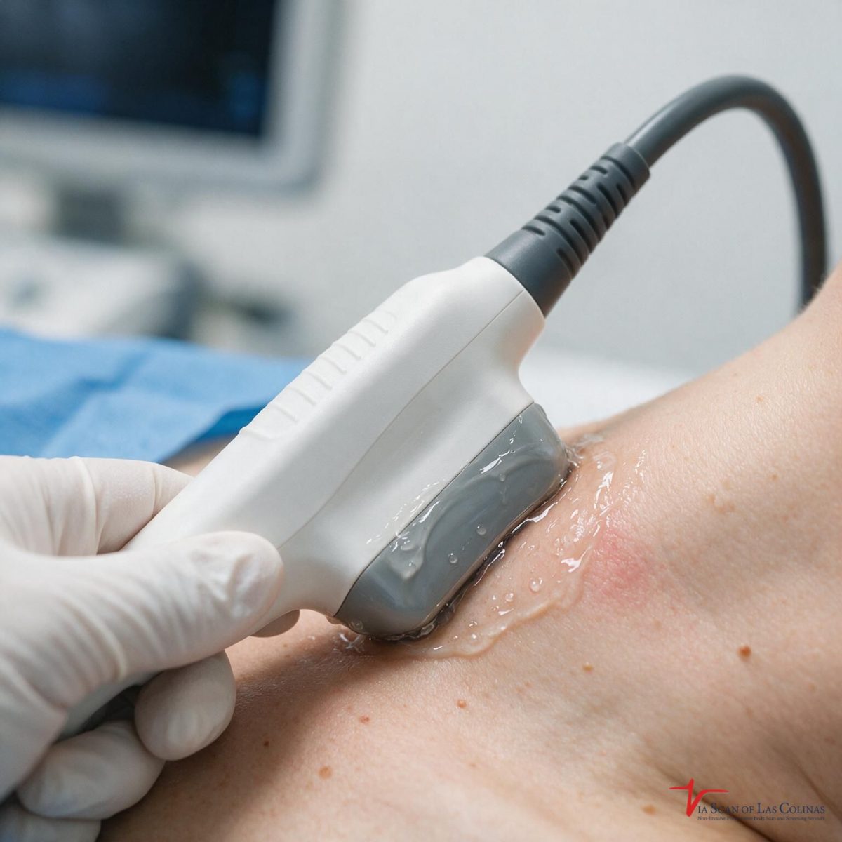
If a neck lump does not go away within the first 2 to 4 weeks, it is a good time to consider medical imaging to evaluate the lump and always sooner rather than later. If you’ve seen a lump on the side of your neck, felt something at the back or had trouble with swallowing and swelling, it’s important to know what’s going on. Neck lumps can be caused by a variety of things, some of which may be harmless, but others will require immediate attention imaging is one of the best ways to distinguish the differences quickly.

What are Neck Lumps and Common Causes of Lumps or Swelling in the Neck?

Neck lumps are any abnormal swelling or mass that is felt or seen in the neck area. Knowing the answer to the question of why does a lump appear in the neck begins with an understanding of the many structures within that small space, which can give rise to a lump for quite different reasons: lymph nodes, salivary glands, thyroid tissue, muscles and blood vessels.

Lymph Nodes, Cysts, Thyroid Nodules, and Other Causes

  • A swollen lymph node is the most common cause of a lump on the side of the neck and is usually a reaction to an infection, usually from a cold, strep throat, or dental abscess, that clears up when the infection is gone.
  • Thyroid nodules are growths in the thyroid tissue on the front of the neck—usually they are benign, but sometimes they need imaging and labwork to determine the significance or nature of the nodule.
  • A cyst, such as a sebaceous or branchial cleft cyst, is a non-cancerous fluid-filled sac that can form anywhere in the neck, at any age, and is usually soft, movable and grows gradually without causing much pain.
  • Neck swelling can also be caused by benign conditions such as lipomas and salivary gland enlargement, although sometimes the appearance of these masses can be mistaken for more serious conditions that need to be clinically assessed by a professional.

When should a neck lump be investigated with medical imaging?

The best question anyone with neck swelling can ask themselves is when to worry about a neck lump. Not all lumps need to be immediately studied by imaging — some traits make professional evaluation truly time sensitive.

Warning signs and factors that may indicate further testing are warranted.

  • A lump that has existed for more than 2-4 weeks and in which imaging and evaluation are warranted, especially if there has not been a recent illness, is a consistent clinical presentation that imaging and evaluation should be performed without delay.
  • A lump on the back of neck or side of neck that is hard, fixed, or is a growing lump is worrying rather than a soft, movable lump  imaging can characterize a lump far more precisely than physical exam.
  • Any neck lump causing difficulty in swallowing, hoarseness, unexplained weight loss or night sweats with swelling significantly increases the clinical concern and warrants immediate medical and imaging work up.
  • Pattern of neck lump cancer symptoms  in adults over 40, a persistent, firm enlargement without pain, especially if the person has a history of smoking or drinking — is always suspicious and should be carefully evaluated with imaging studies.

Which Imaging Tests Are Used To Evaluate Neck Lumps?

There are several ways to assess a neck lump and the most appropriate one will depend on what is suspected to be the cause of the lump, where the lump is located and how much additional information is required apart from what is found on physical examination.

How Ultrasound compares to CT Scans and MRI.

The first imaging test used for a lump on the side of the neck is usually ultrasound, which is quick, gives no radiation, and can be used to help determine the characteristics of lymph nodes and distinguish solid masses from fluid-filled cysts.

  • When cancer symptoms are suspected in a neck lump and the involvement is suspected to be deep, CT scans can give detailed cross-sectional imaging—very useful for this purpose.
  • MRI is excellent for soft tissues, does not use radiation and is preferred in the imaging of deeper neck compartments and salivary glands where high resolution differentiation of tissues is important.
  • PET-CT may be performed when malignancy is suspected, to find out if the tissue is growing and for better detection of any other areas of involvement than the main lump.

How Can Medical Imaging Help Detect Potentially Serious Neck Conditions Early?

Medical imaging can detect serious neck problems early because it shows abnormalities that cannot be seen on a physical exam, such as small, deep masses, early thyroid changes, and enlarged lymph nodes that don’t yet cause surface swelling. Imaging data adds to the clinical picture to make decisions about when to be concerned about a neck lump clearer. Early imaging is a valuable tool for persistent or concerning swelling, as there is often a definitive answer as to what is causing a lump in the neck.

If you prefer to see things in detail and from an anticipatory perspective, ViaScan of Las Colinas provides non-invasive CT scans and whole-body scans that scan multiple regions of the body at once. A whole-body scan can detect abnormal findings in the neck along with other internal abnormalities, aiding in early, informed discussions with your health care provider. If you have a neck lump and are having trouble swallowing or have a lump on the back of your neck – talk to a licensed professional before adding preventive imaging to your health plan.

Choose Our Wellness Body Scan

Early Detection Saves Lives!

    • Accurate
    • Quick Result
    • Affordable

Conclusion

Neck lumps occur regularly and are generally non-cancerous but if the lump is persistent or accompanied by symptoms, it is important to seek professional advice as soon as possible. ViaScan of Las Colinas provides professional CT scan and whole body scan services to people with a proactive approach towards health awareness, knowing early is better than knowing later. Seek professional assistance early; early assessment always results in better outcomes.


Lower-Left-Abdominal-Pain-Causes-1200x675.jpg
29/Jun/2026

The lower left quadrant of the abdomen can be affected by a multitude of illnesses, from a flare-up of the gut to an emergency. It is where the descending colon, left kidney, left ureter and for females, left ovary and fallopian tube are located. The pain here is a message from your body to “pay attention”. This blog provides you with a step-by-step analysis of when to use imaging and warning signs and symptoms.

What are the most common causes of lower left abdominal pain?

The lower left abdominal pain is usually due to digestive, urinary, reproductive or musculoskeletal issues. The nature of the pain, its onset, and its accompanying symptoms give valuable hints to the involved system.

Gastroenterological, urinary, reproductive, musculoskeletal disorders.

  • Diverticulitis — Inflammation of the pouches in the colon wall brings on sharp localized pain in the lower left quadrant of the abdomen, sometimes with fever, nausea and altered bowel movements, which are a warning sign of a digestive disorder that must be evaluated by a medical professional.
  • Lower left pain radiating up to groin or back, often with urinary frequency or pain on urination and/or blood in urine, may be due to kidney stones or UTI (kidney infection) that are moving through the left ureter.
  • For females, the left ovary may cause chronic/mild pain during their periods, or be the cause of sudden sharp pain that may be due to a left ovarian cyst, endometriosis or even ovarian torsion, all demanding varying degrees of medical urgency and evaluation.
  • Physical effort or weakened abdominal wall, muscle strain or hernia — Musculoskeletal pain from physical exertion or a weak abdominal wall which gets worse with exercise or pressure but no digestive or urinary symptoms.

What could be the serious condition if the lower left abdominal pain has symptoms?

Some symptoms in combination with lower left abdominal pain indicate that there is a serious problem that requires immediate medical attention.

Warning Signs that should not be ignored:

  • If there is fever with lower left pain, then it is likely that there is some infection or inflammation such as diverticulitis or a kidney infection which needs professional evaluation and should not be treated at home or delayed beyond a few hours.
  • If left abdominal discomfort is accompanied by blood in the stool or urine, this is a sign that you are suffering from a digestive or urinary issue that needs medical imaging to determine the exact cause and rule out serious underlying problems.
  • If the pain is sudden, severe and quickly getting worse, it could be a surgical emergency like a ruptured cyst, ovarian torsion or perforated bowel and care should be sought immediately without waiting for symptoms to improve.
  • If the vomiting continues or gas is seen to be trapped and the infant does not defecate, this may indicate an intestinal blockage which should be investigated immediately and should not be ignored for longer than several hours.

When should you seek medical attention for lower left abdominal pain?

If pain is sudden, severe or worsening quickly, get emergency medical help right away. A scheduled appointment is appropriate when the discomfort is mild and improving and occurs intermittently. The following table will help you make a decision.

If any doubts, please get medical evaluations. Persistent or new or worsening lower left abdominal pain is a sign to seek medical attention.

Emergency Symptoms vs. Non-Urgent Discomfort

Seek Emergency Care Schedule an Appointment
Sudden or severe pain Mild pain that comes and goes
Fever above 101°F Dull ache without other symptoms
Blood in stool or urine Bloating or gas-related discomfort
Rigid or tender abdomen Pain that improves with rest
Dizziness or rapid pulse Cyclical pain tied to menstrual cycle

When Is Imaging Recommended for Lower Left Abdominal Pain?

Imaging is advised when there is no physical or laboratory finding to determine the etiology or when a serious condition must be quickly established or excluded.

What role do CT Scans and Other Tests Play in Finding the Cause?

  • A CT scan abdomen for lower left pain offers detailed cross-sectional imaging of abdominal organs, bowel, and surrounding tissue, and is one of the most informative and rapid tools available for the evaluation of the acute abdomen.
  • A contrast CT scan abdomen and pelvis will show inflammation, blood vessels and boundaries between organs more clearly and is especially helpful to confirm masses that are not seen on other imaging, diverticulitis or kidney stones.
  • Medical imaging for left abdominal discomfort that is chronic may include an MRI abdomen for chronic pain evaluation — preferred when soft tissue detail is required and repeated radiation exposure is a clinical consideration for the patient.
  • When a woman has reproductive problems, ultrasound is usually the first procedure ordered before a contrast CT scan abdomen and pelvis or an MRI abdomen is ordered, and when the initial ultrasound results are not conclusive, a contrast CT scan abdomen and pelvis or MRI abdomen is ordered next to evaluate chronic pain.

At ViaScan of Las Colinas, we provide a non-invasive preventive imaging service, which includes a whole-body scan that covers the abdomen thoroughly, for those who experience regular abdominal pain and are interested in a more comprehensive examination of what is going on inside. If there are cardiovascular issues in addition to abdominal symptoms, a heart scan can offer comprehensive information about the heart in a preventive evaluation. These are awareness tools, NOT emergency care, and are best used in union with instructions from a licensed health care provider.

Choose Our Preventive Heart Scan

Early Detection Saves Lives!

    • Accurate
    • Quick Result
    • Affordable

Conclusion

Lower left abdominal pain is not something to take a stab at. The causes can be manageable or urgent; the appropriate action will depend on your symptoms. For anything new, persisting, or getting worse or anyone who wants to make proactive health choices, ViaScan of Las Colinas offer professional preventive imaging that can help with informed, early health choices.