Do you have an appointment for a CT scan, and you’re unsure how long it will take? The duration of the procedure and the experience of being inside the machine are the major concerns of many people. The best part is that the CT scans are much faster than most people think. This blog discusses the average turnaround time and how ViaScan makes the process quick and comfortable.
As a medical professional, I work with patients daily, and most of them have numerous questions about CT scans. The most common concerns are the duration of their stay, whether they will be at ease, and what happens if something goes wrong. All these are justifiable questions. The following are the processes involved in the CT, so you get to know what to really expect when you get to Via scan.
How Much Time does a standard CT Scan take?
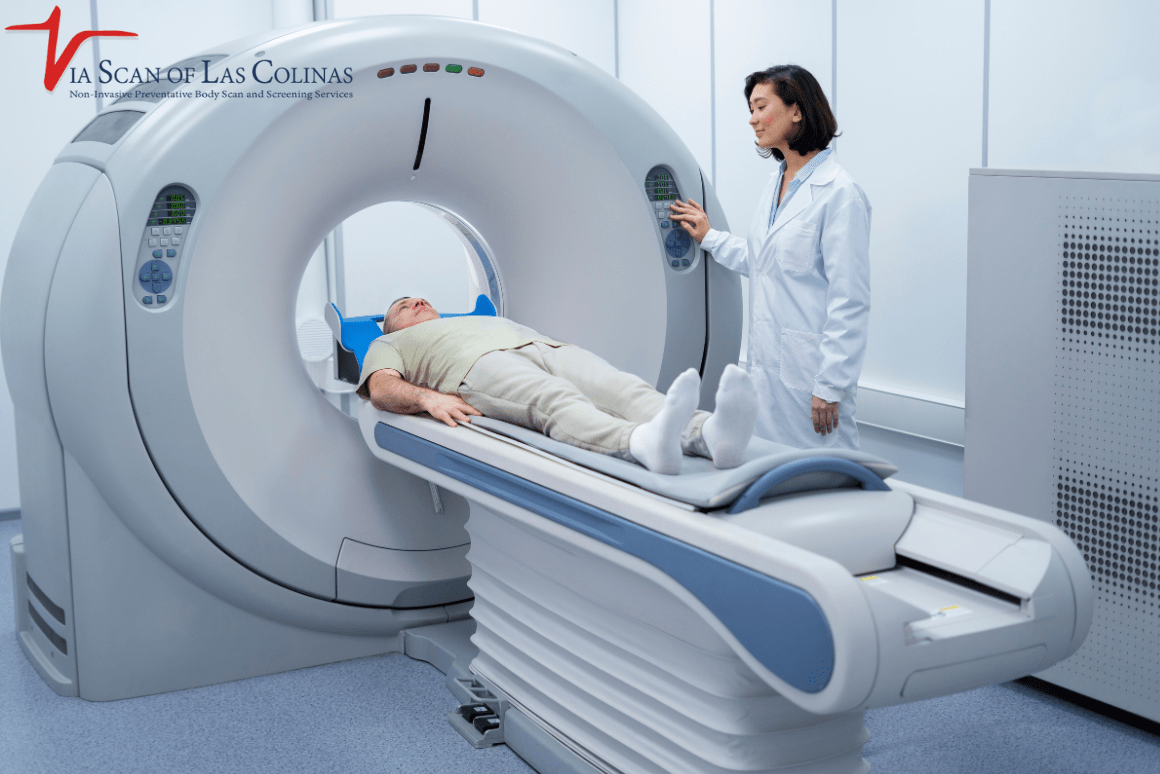
- Common CT is approximately fifteen to thirty minutes to complete. The actual scanning, which is the time taken in the machine, takes only five to ten minutes.
- Prior to taking the scan, medical personnel will admit you, ask health questions, and prepare you. Following the scan, you will have to wait a couple of minutes while we ensure that the images are clear and of good quality.
- ViaScan trains its staff to work fast and efficiently. We realize that the patients are busy, and that is why we ensure the CT scan process takes as little time as possible and provide the best images to your physicians.
- At ViaScan, we have advanced technology and qualified personnel who ensure everything runs smoothly and quickly.
Is a CT scan Longer When the vital organs are involved?
Various parts of the body, varied scan time:
- Head scan: Five to ten minutes of real-time scanning.
- Scan of the neck: Approximately eight to ten minutes.
- Chest scan: Approximately ten or fifteen minutes.
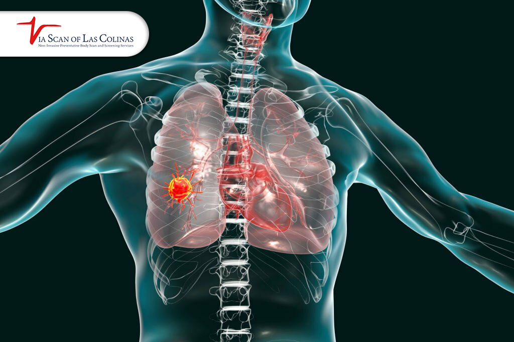
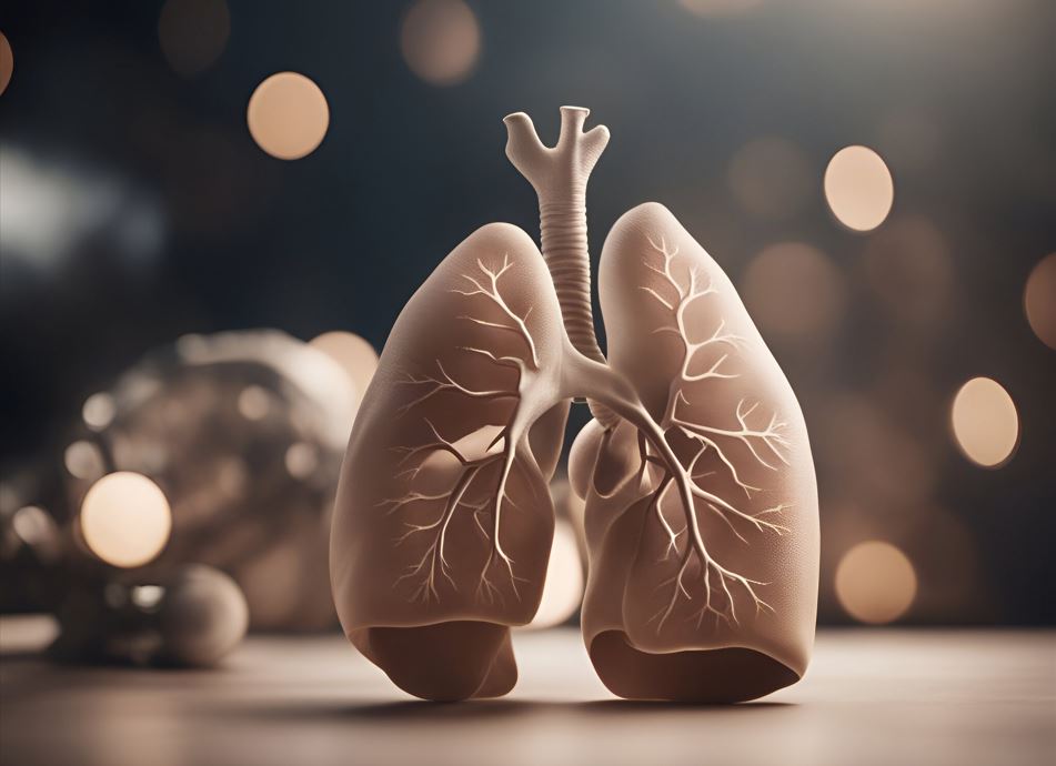
- Heart scan and lung scan: Ten to fifteen minutes
- Scanning of the abdomen: Approximately ten to fifteen minutes.
- Pelvis scan: approximately ten to fifteen minutes.
- Spine scan: Approximately ten to twelve minutes.
- Whole body scan: fifteen to twenty minutes
Why times vary:
- A full body scan takes more time to image.
- Additional angles and views are required in some scans.
- The imaging protocols of various body parts vary.
At ViaScan, we excel in any form of scans, such as cardiac, lung, and full body scans. We scan you in the shortest amount of time using the least amount of scanning, so comfortably.
What factors affect the time taken by a CT scan?
There are various ways in which a CT scan may take longer than expected.
- The scan will have to be repeated a few times if you are not still, which will increase the time.
- When the doctor orders additional images or different angles, that consumes time.
- Sometimes it takes longer to calm down the patient if he feels nervous or uncomfortable.
- In case a contrast dye is involved, we must wait, you see, until the contrast dye has circulated in your body.
- Machine fine-tunings or technical problems could cause additional time.
- A heavy working schedule with several patients per day can cause a minor wait.
We attempt to reduce delays at ViaScan. Our employees are trained to help you feel calm and still. We have the latest machinery that is highly dependable. If the scan involves contrast dye, we will notify you in advance to avoid any unpleasant surprises regarding the CT scan duration.
How Does the Use of Contrast Dye Affect the Total Time of the Scan?
Aspect |
CT scan Without Contrast |
CT scan With Contrast |
| Time for setup | Five minutes | Ten to fifteen minutes |
| Reason for extra time | No IV line needed | Need to insert an IV line in the arm |
| Waiting time before scan | None or minimal | Five to ten minutes for the yeast to work |
| Actual scanning time | Five to ten minutes | Five to ten minutes |
| Waiting time after scan | None or minimal | Five to ten minutes (observation) |
| Total visit time | Fifteen to thirty minutes | Twenty to forty minutes |
| Image quality | Good for most areas | Excellent for soft tissues and blood vessels |
| Warmth feeling | Not applicable | You may feel warm (normal) |
| Nausea risk | Very low | Very low (allergies are more common) |
| Who receives contrast | People with specific health needs | Doctors order when better images are needed |
| Cost difference | Standard price | Slightly higher due to contrast material |
Contrast dye enables your doctor to have a better picture of the organs. At ViaScan, your comfort is a priority for us. Only the FDA-approved safe dyes are utilized. In case of allergy or medical-related issues, inform us during your booking, and we will ensure to take the necessary measures. The awareness of how long does a CT scan take with contrast will enable you to be prepared.
The best choice of CT imaging system is ViaScan.
ViaScan believes that medical imaging must become quick, comfortable, and precise. Our machines are up to date with industry standards, run more rapidly, and produce clear images compared to older models. It provides faster scans at better quality for your physicians. Our employees are qualified radiologists and technicians. They train to ensure you are safe and at ease.
We elaborate it to you using plain words so that you are not left guessing what is going on. There are too many types of scans, such as heart, lung, and whole body scans. You have a few scans to do, or you may need a longer examination of the entire body. We are professionals with the machines to assist you with your scans.
Choose Our Preventive CT Scan
Early Detection Saves Lives!
-
- Accurate
- Quick Result
- Affordable
Conclusion
In case you have a CT scan planned, this is what you should anticipate. An average scan would last around fifteen to thirty minutes, but the actual scanning would last only five minutes or fewer. The body parts take varied durations. Contrast dye provides doctors with far more information. It is a non-invasive and non-painful procedure. At ViaScan, we maintain a low scan time, ensuring high-quality images. Like the heart and lung scans, we provide whole-body scanning services using the same fast and accurate approach. Professional assistance on your medical imaging needs. Your health is important, and we are the ones to provide you with the answers.