Aortic-Aneurysm-in-females-symptoms.jpg
24/Mar/2026

Aortic aneurysms, or life-threatening dilations of the largest vessel in the human body, frequently go undiagnosed, which makes them especially dangerous in women because they might not notice signs until a medical health emergency takes place. Knowledge about the symptoms of aortic aneurysm in females makes women recognize possible issues at an earlier stage when preventive screening and treatment can be used to avoid life-threatening ruptures. Although aneurysms develop more frequently in men, women may present with less typical symptoms compared to men and women should be aware of them to detect them as early as possible. 

What are typical symptoms of an aortic aneurysm in females that can not be overlooked?

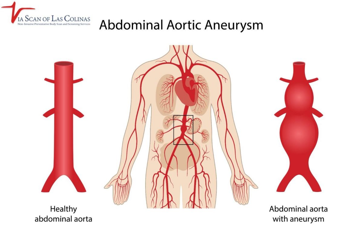
A significant number of aortic aneurysms in females grow to large sizes without any obvious symptoms before rupture. But when the symptoms occur, women should not overlook them. The typical symptoms are chest, abdominal, or back pains that are not relieved by rest. There are women who experience the sensation of pulsing around the belly button or abdominal fullness. Sudden intense pain, dizziness, rapid heart rate, or shortness of breath are the signs of possible rupture and immediate emergency assistance is needed. Awareness of aortic aneurysm symptoms in females is essential for early diagnosis and treatment of the warning signs and taking immediate medical care involving diagnostic tests.

Early Warning Signs in Women

Persistent pain of an abnormal type: Pain in the chest, upper back, or abdomen that does not come and go, that is a constant deep pain, not the sharp pain associated with normal muscular pain.

Pulsating sensation: The sensation of a throbbing or a beating in the abdomen, especially around the region of the belly button, which beats in sync with your heartbeat, often noticed when lying down.

What is the difference between the symptoms of an aortic aneurysm in women and men?

Studies indicate that women have various and even less obvious symptoms of aortic aneurysms than men, which may delay diagnosis and increase the likelihood of rupture. Women tend to manifest more with non-characteristic symptoms not immediately identified as heart difficulties such as stomachache that is often mistaken for digestive issues, back pain, which may be attributed to musculoskeletal disorders, or non-specific chest pains that may be misinterpreted as anxiety. The aneurysm also develops in women at a later age than men and in most cases with smaller body sizes that is, the aneurysm may burst at less diameter in women than in men, thus early preventive screening is of utmost importance, particularly to women.

Gender Differences in Symptoms.

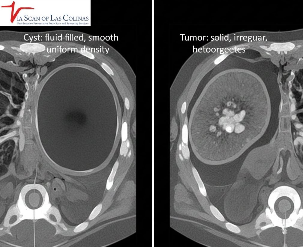
Women are more likely to report abdominal pain and nausea than men when they have aortic aneurysms, which is frequently regarded as a symptom of gastrointestinal disorders and not cardiovascular ones. The patients with female gender report back pain more frequently in comparison with male patients, especially when thoracic aortic aneurysms occur in the upper part of the aorta. Also, rupture rate among women at smaller size of aneurysms is higher than that of men, so symptoms can manifest themselves at relatively small aneurysms, and diagnostic imaging tests should be considered by CT scan or other sophisticated imaging systems.

What is the experience of pain due to an aortic aneurysm in women?

The aortic aneurysm pain in women depends on the site of the aneurysm and the size of the aneurysm and the stability or enlargement of the aneurysm. Women characterize the sensation in other ways, yet, there are regular patterns, which assist in the detection of possible aneurysms that may be evaluated by medical professionals. The pain is also deep, constant and chronic as opposed to acute or stabbing. It does not usually react to over-the-counter painkillers and can progressively increase the size of the aneurysm in weeks or months.

Patterns of Chest, Back or Abdominal Pain.

  • Thoracic aortic aneurysm pain: Deep pain in the chest or upper back, which can extend between the shoulder blades, can be characterized as a tearing or ripping feeling, in case the aneurysm is dissecting or growing at an accelerated pace.
  • Pains of abdominal aortic aneurysm: Pain in the lower back or belly, which is steady and deep, at times with a pulsating sensation around the belly button, or even like the belly was being pressed full.
  • Pain characteristics: Unlike muscle pains which can be relieved by a change of position or rest, a person will always experience the pain with an aneurysm, and over time it might be more intense but it does not disappear entirely but is noted as a deep pain that does not disappear altogether.

Are there women who may have an aortic aneurysm without any symptoms?

Yes, and this is what makes aortic aneurysms so dangerous, most of them grow totally silent and do not produce any symptoms, when they are very large or rupture. The aortic aneurysms in many women are accidentally found when an imaging is performed due to other medical causes, and the women had been unaware of the existence of the aneurysm. This silent development means that women who are at risk, including those with high blood pressure, have a history of smoking, or there are aneurysms in the family, or have a history of connective tissue disorders should seek preventive screening despite the absence of symptoms to ensure the identification of aneurysms at an early stage so that they can be monitored thus preventing rupture.

Significance of Screening on a Periodic basis.

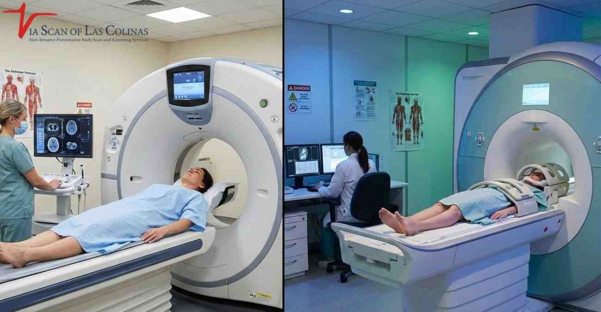
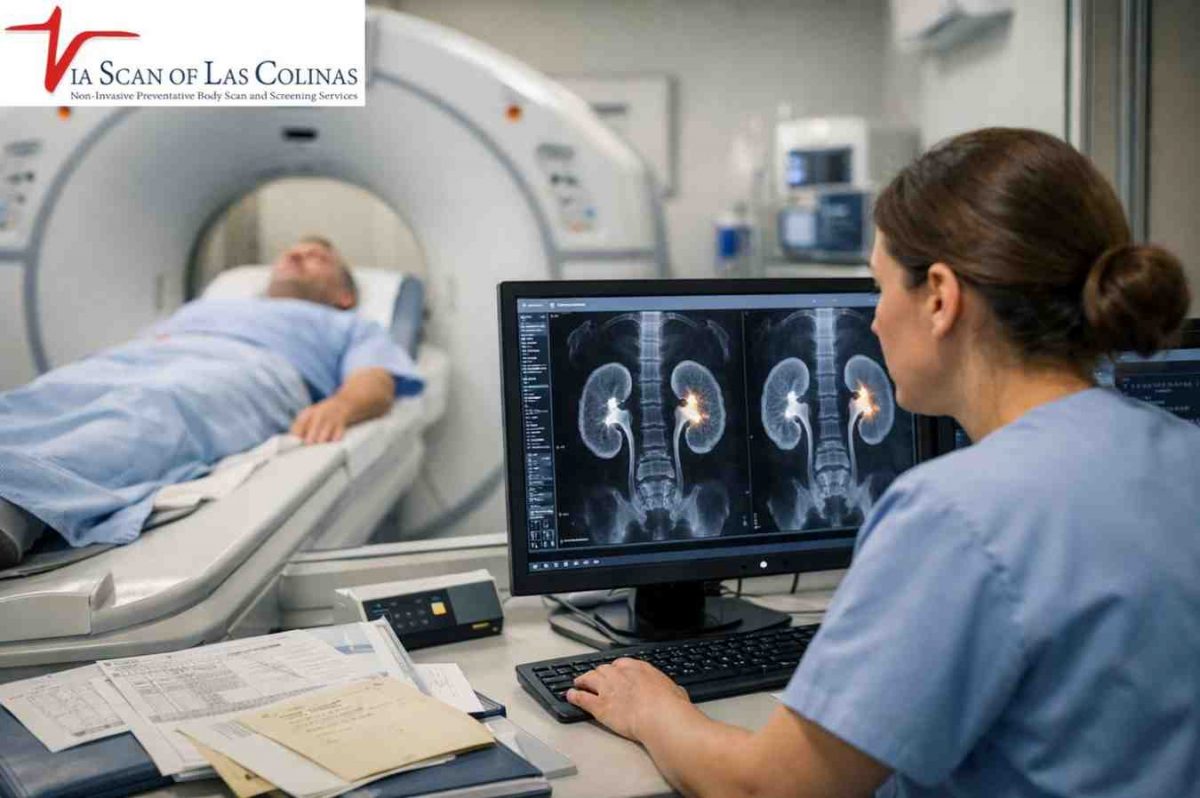
  • Women with history of aortic aneurysms in their family, women who have smoked, women with high blood pressure, women with atherosclerosis, and women with connective tissue disorders are the most likely beneficiaries of screening. The CT scan services offered by ViaScan offer high-quality aortic imaging to identify aneurysms in women even before they start to appear.
  • Monitoring: Preventive screening helps to monitor small aneurysms which can be followed up over time with periodic imaging and medical treatment can be initiated before it is deemed large enough to cause harm and necessitate emergency treatment.
  • Benefits of whole-body screening: Comprehensive whole-body scanning, besides aortic aneurysms, also identifies other silent cardiovascular and health conditions, which offer full preventive health care to women who prioritize wellness as their first line of priority.

Choose Our Preventive CT Scan

Early Detection Saves Lives!

    • Accurate
    • Quick Result
    • Affordable

Conclusion

Learning the symptoms of aortic aneurysm in female and seeking preventive screening will literally save your life by preventing dangerous aneurysms before they burst. In Irving, TX, ViaScan offer the most advanced CT scan and complete whole body scan which can discover aortic aneurysms and other cardiovascular diseases during preventive health screening.


Advanced-Kidney-Cancer-Diagnostic-Imaging-_-ViaScan-1200x800.jpg
16/Mar/2026

Kidney cancer is relatively common among adult cancers and is usually silent and does not produce any visible indicators of its presence at early stages, and it is difficult to detect without effective medical examination and radiography. Learning the process involved in diagnosing and identifying kidney cancer makes you appreciate the need to get regular health checkups particularly in cases where you are at risk of developing such risk factors as smoking, obesity, high blood pressure, or even family history. Timely diagnosis of kidney cancer is a significant aspect in the treatment process and that is why the use of sophisticated imaging is such an important factor in ensuring that kidney abnormalities are detected in good time to prevent them. 

What are the methods through which physicians detect kidney cancer at early and advanced stages?

Diagnosis of kidney cancer entails a review of the medical history, physical examination, laboratory tests, as well as advanced imaging studies, which present kidney abnormalities. Numerous cancers of the kidney are incidentally identified at the time of imaging to show some other cause as an early tumor is not likely to have symptoms. Whenever the kidney cancer symptoms are observed, i.e., blood in the urine, chronic back or side pain, unexplained weight loss, or fatigue, the symptoms may indicate a more advanced disease, which needs urgent examination and prolonged diagnostic tests to see the stage of the cancer and the available treatment options.

Primary Medical Examination and Signs of Kidney Cancer.

At the time of the initial examination, physicians enquire about symptoms of kidney cancer such as blood in urine (hematuria), chronic pain in the back or lower abdominal side below the ribs, unexplained weight loss, fever, fatigue, or palpation, an abdominal mass. A swelling or an abdominal mass may be detected on physical examination but this is not common in early stage cancer of the kidney. The majority of kidney cancers are silent and are detected accidentally in abdominal imaging due to other conditions at an early phase, and therefore, screening of health condition and diagnostic imaging is important to detect the risk factors.

Diagnostic Tests used for Confirmation

After the suspicion of kidney cancer, physicians test to diagnose and identify the extent of the cancer. Several imaging procedures such as CT scan of kidney cancer, MRI, and kidney ultrasound test give detailed images of the kidney structures showing the size, location and characteristics of tumors. Blood tests and urine tests evaluate kidney functioning and detect abnormalities that may be the indication of cancer. In certain cases, tissue biopsy can be done so as to study the cells under a microscope, but imaging can in most instances of kidney cancer suffice to make diagnosis and implement treatment.

What are the commonly used imaging tests to identify kidney cancer?

The modern imaging is the new kidney cancer diagnosis method where one can view a detailed kidney structure that depicts the presence of masses, tumors, and other abnormalities that are not directly seen or detected by the physical examination and blood test only. Kidney cancer CT scan is the most widely and valid imaging technique but MRI and kidney ultrasound test offer useful diagnostic information in certain circumstances. The professional CT scan services offered by ViaScan provide high quality imaging that are required to detect renal cancer and fully assess the kidney.

CT Scans, MRI, and Ultrasound

  • CT scan of kidney cancer: CT scans give excellent visual representations of cross-sectional pictures of kidney tumors with good level of clarity – the CT scan is enhanced with contrast that shows the blood supply to the tumor and differentiates between cancerous and benign cysts.
  • MRI imaging: Magnetic resonance imaging has better soft tissue contrast that can be used in cases where CT findings are inconclusive or in patients who cannot be administered CT contrast dye especially when determining the spread of tumors into blood vessels.
  • Kidney ultrasound test: This is an image test that involves the use of sound waves to provide real time images that help differentiate fluid filled cysts from solid masses – this is usually the first test to be performed on the kidneys when the abnormalities are suspected but not as detailed as CT or MRI.

The way Imaging Discovers Tumors in the Kidneys.

  • Mass identification: Imaging clearly reveals abnormal growth or malformation in kidney tissue in that it distinguishes between solid tumors and fluid-filled benign cysts that do not necessitate treatment.
  • Size and location mapping: The detailed images indicate the exact size of the tumor, its location in the kidney, and its connection with the surrounding organs – this is very important when treating the tumor.
  • Staging information: Imaging defines where kidney cancer has spread outside of the kidney onto the lymph nodes, blood vessels, or other organs nearby to the kidney – necessary to identify the stage of the cancer, and the way to treat it. Imaging findings can also help physicians understand how fast kidney cancer spreads and whether nearby organs or lymph nodes are already involved.

How long does it take to die from kidney cancer

The progression of kidney cancer varies greatly depending on the stage at diagnosis, tumor aggressiveness, and overall patient’s health condition. Early stage kidney cancers that are detected incidentally often have a much slower progression, and timely treatment can significantly prolong survival. Developed kidney cancer, particularly stage 4, can spread quickly to distant organs such as the lungs or bones, which may shorten life expectancy if untreated.

It is more important to note that each patient’s course is unique and survival depends on factors such as age, kidney function, response to treatment, and the presence of other health conditions. Regular monitoring and early intervention are key to managing the disease and improving outcomes.

What is the use of blood and urine tests in the diagnosis of kidney cancer?

Although the imaging can give a visual confirmation of kidney masses, blood and urine tests can be used to give deepening diagnostic information on the functioning of kidneys and health condition overall. These laboratory tests do not actually diagnose kidney cancer but assist in the diagnosis of abnormalities that need further investigation and give baseline health data that will be critical in the planning of treatment. Complete diagnostic assessment of kidney cancer requires evaluation by use of imaging as well as laboratory tests which are best done at an early stage.

Diagnosis of Abnormal Function of the Kidney.

  • Blood urea nitrogen (BUN) and creatinine: An increase of these values reflects kidney dysfunction which could be as a result of cancer of the kidney tissue.
  • Complete blood count (CBC): Discloses anemia, which is a frequent symptom of kidney cancer, or the presence of high red blood cell counts (polycythemia) that is occasionally caused by kidney cancer.
  • Liver function tests: The abnormal results can give an indication that kidney cancer has metastasized to the liver, although this is not the main task of these tests.

Evidence for Continued Testing.

  • Urinalysis: Reveals blood in urine (hematuria) – a frequent symptom of kidney cancer that would then lead to imaging to establish the source of bleeding.
  • Blood calcium level: Increased calcium (hypercalcemia) occasionally points to high grade kidney cancer that causes hormones that influence calcium metabolism.
  • Lactate dehydrogenase (LDH): High levels could be an indication of aggressive kidney cancer, but this is a nonspecific marker that needs to be confirmed by imaging.

Choose Our Preventive CT Scan

Early Detection Saves Lives!

    • Accurate
    • Quick Result
    • Affordable

Conclusion

Early diagnosis of kidney cancer enhances success rates and survival with a high degree of success. We offer specialized CT scan services and whole-body scan options that are at par with those offered in ViaScan, Irving, TX, to assist in diagnostic services of renal cancer in infancy and renal health checks using diagnostic imaging services.


Abdominal-Aortic-Aneurysm-Symptoms-1200x792.jpg
09/Mar/2026

The aorta is the largest blood vessel in your body; it is the one that carries oxygen-rich blood to the other parts of your body. An abdominal aortic aneurysm happens when a part of the aorta in your abdomen is weak and weakens and bulges out like a balloon. This is a severe condition as the aneurysms may develop without any symptoms, and they may burst without warning with lethal effects. Understanding the symptoms of abdominal aortic aneurysms, even the slightest signs, can actually make the difference between saving your life and losing it, as it is time to get medical attention before the rupture happens.

A screening of abdominal aortic aneurysms during the early stages of their development can enable medical personnel to examine aneurysms and prevent the occurrence of emergencies. 

What are the initial symptoms of an abdominal aortic aneurysm that human beings should never overlook?

Numerous abdominal aortic aneurysms will never present any symptoms, hence the title of silent killers. Nevertheless, the symptoms should not be disregarded once they come. The symptoms that are common in the early stages consist of some persistent or aching pain located in your abdomen or lower back, a feeling of pressure or fullness in the abdomen, or a pulsating aching feeling below your belly button when lying down.

Some experience a persistent pain that does not resolve in the abdomen or the back. Although these symptoms may be caused by other factors, their occurrence particularly in individuals with risk factors such as smoking, high blood pressure, or a history of family members will necessitate professional examination and screening of abdominal aortic aneurysm to eliminate this life-threatening disease.

Warning Signs of an Enlarging Aneurysm

  • Pulsating abdominal mass: To be able to feel a rhythmic pulsing of your abdomen, especially around the belly button, that beats with your heartbeat – this can be a sign of an enlarging aneurysm.
  • Unidentified lower back pain: Persistent deep pain in the lower back that cannot be identified as having any specific cause, which does not lessen even when one rests or resorts to customary pain management methods.

What does an abdominal aortic aneurysm pain feel like?

In the cases of an abdominal aortic aneurysm, patients tend to complain of a deep, nagging ache as opposed to sharp or stabbing pain. The pain is normally located in the lower back or the abdomen and might present as a continuous, gnashing pain that does not entirely resolve.

Other individuals say that they experience a sensation of something pressing or pulling in their stomach. As opposed to the infrarenal abdominal aortic aneurysm pain which occurs below the kidneys, an ascending aortic aneurysm produces other symptoms that occur in the chest and not the abdomen. The difficulty lies in the fact that the pain of an aneurysm is somewhat nonspecific and may be confused with muscle aches, stomach problems, or kidney diseases. This is why the diagnostic tool of professional imaging should be taken into consideration to identify the problem properly.

Is it possible that an abdominal aortic aneurysm can occur without any signs?

Yes, and that is why abdominal aortic aneurysms are so dangerous, most of them arise absolutely without any symptoms, and only when they are very large or rupture. The majority of small to moderate-sized aneurysms do not lead to any symptoms at all, and hence screening is essential in people who are at risk. The most common type of aneurysm that may develop below the arteries that serve the kidneys is called the infrarenal abdominal aortic aneurysm and may take years to develop, unless screening is performed. This is a silent course and, therefore, the individuals are usually unaware that something is amiss until a medical crisis occurs or an aneurysm is found during imaging done due to other causes.

Significance of Preventive Screening

Early intervention saves life: a CT scan imaging of abdominal aortic aneurysms can diagnose a aneurysm before the onset of symptoms , which means that the aneurysms can be monitored and treated before the risk of rupture becomes lifethreatening.

The most benefited people are men aged 65 to 75 who have smoked in their lives, a person with a family history of aortic aneurysms,  and individuals with atherosclerosis should have a discussion with healthcare providers about screening with the ViaScan whole-body scan.

What are the Symptoms that Can be Observed in Case of an Abdominal Aortic Aneurysm that Starts Rupturing?

Rupturing an abdominal aortic aneurysm is a healthcare emergency that can’t be attended to without emergency care. The symptoms of rupture or impending rupture are abrupt, and dramatic and quite unlike the insidious symptoms of stable aneurysms.  these indicators of emergencies can be the only difference between life and death when they are known, and a call is made to 911. Do not wait or attempt to find out whether they improve; immediate medical help is critical to survival.

Emergency Warning Signs

  • Severe abdominal or back pain:  Sudden, or tearing or ripping, abdominal or back pain that occurs suddenly and is unlike anything one has experienced before.
  • Rapid heart rate, lightheadedness, weakness, or fainting; your heart is racing, you are feeling lightheaded, weak, or faint as if someone were bleeding inside you.
  • Clammy Skin and Unconsciousness : Sweat, Pallor or Bluish skin color, confusion or passing out, these are symptoms of shock caused by serious internal bleeding that has to be treated as an emergency.

The Importance of Early Detection.

Since the majority of the abdominal aortic aneurysms are asymptomatic until they attain a dangerous size or rupture, proactive screening is of paramount importance to high-risk patients. CT scan imaging provides clear and detailed images of the aorta, measuring the size of aneurysms more precisely and assisting medical professionals in making appropriate contemplations of which strategies to follow during monitoring or intervention. In contrast to ascending aortic aneurysm, which involves the chest part of the aorta, infrarenal abdominal aortic aneurysm diagnosis requires abdominal imaging of the lower part of the aorta, which is well seen.

Choose Our Preventive CT Scan

Early Detection Saves Lives!

    • Accurate
    • Quick Result
    • Affordable

Conclusion

This silent condition can be detected at an early stage with the help of abdominal aortic aneurysm screening before it becomes fatal. We offer progressive CT  and heart scan services, and full body scan services in ViaScan, located in Irving, TX, which help detect abdominal aortic aneurysms and other severe diseases during preventive screening of our health conditions.


whole-body-scan-ViaScan-1200x750.jpg
02/Mar/2026

After a good night’s sleep, you wake up feeling pained in the back and ribs, and your day starts off frustrating. You are no longer refreshed and ready; you feel stiff, sore, and wonder why you wake up with a sore body after hours of sleep. Rib pain in the morning or rib cage pain after sleep is a common symptom in many individuals, and knowing the causes will assist you in concluding whether it is a mere sleep-related or a medical problem. Although it is normal to have temporary muscle stiffness after sleep, pain that is persistent or increasing is an indication of underlying conditions that require professional care. 

Why do I experience pain in my back and ribs upon waking up in the morning?

Back and rib pain in the mornings occurs because of different causes, including the mere problems with sleeping positions and some more severe medical disorders. Mattress support is poor, thus causing your spine to bow down in an unnatural manner resulting in pressure points to cause pain in the rib cage after you wake up. Natural issues such as arthritis, muscle injuries, or back complications tend to be more painful in the morning after rest. Whether it is normal to find the rib sore after waking up is a question that would depend on frequency and intensity, infrequent and mild pain is a common feature, but when pain is frequent and severe, it would be appropriate to seek the services of a professional.

Daily Habits That Lead to Overnight Pain

  • Sedentary life: Sitting in the same place hours results in stiffening of both muscles and joints and results in temporary pain in the ribs at mornings and back pain which often subsides with movement during the day.
  • Imperfect positioning of sleep: Sleep posture that twists the spine, intercostal muscle strain, or joint misalignment puts pressure on the thoracic cage, which accumulates when one sleeps, leading to pain on awakening which can be found till the muscles relax and restart positioning.

When Mornings Stiffness Is a Red Flag.

  • Constant intense pain: Morning pain that does not dissipate in the first 30 minutes of waking, or gets worse throughout the weeks or disrupts daily living might be a sign of chronic back pain causes that need medical imaging test.
  • Companion symptoms: Pain with fever, unexplainable weight loss, shortness of breath, or weakness is an indication of severe disorders that need immediate medical evaluation involving diagnostic tests such as CT scans.

Am I experiencing back and rib pain because of my sleeping position?

Of course, one of the most frequent causes of rib cage pains after sleeping and morning back discomfort is the sleeping position. Stomach sleeping causes your neck to bend sideways hours, the muscles of the neck and upper back work hard and the rib cage is positioned at an awkward angle. Sleeping on the side without proper support enables the top shoulder to roll forward, squeezing the rib cage and forming an imbalance in the body structure.

Even the back sleeping position will be troublesome in case your pillow is either too high or too low, thus forcing your back into unnatural positions. Being aware of what positions cause strain will enable you to make the necessary adjustments that could decrease the level of muscle tightness following sleep and prevents the occurrence of rib pains in the mornings before it occurs.

Is my mattress/pillow causing any back and rib pain in the morning?

Surely, that is true- your mattress and pillow make a huge difference in you waking up pain-free or achy. A mattress that is way too soft will result in an uneven sinking of your body, which results in misalignment of your spine and pressure points that will lead to you feeling some pain after sleep in your rib cage. A mattress that is too firm does not provide adequate cushioning of pressure areas, causing pain in certain body parts such as shoulders and hips and extending the pain to other parts such as ribs and back. The old mattresses become soft and form sagging areas which make your back bend in unhealthy postures when sleeping. Likewise, the pillows which are not suitable to your sleeping position do not provide the neck and spine with the right position during the night.

The selection of the Pillow and Firmness.

  • Back sleepers: require medium-firm mattresses with medium pillow height to maintain the natural curve of the neck without either pushing the head too far forward or letting it fall back.
  • Side sleepers: prefer medium to medium-hard mattresses with firmer and higher pillows to fill the space between shoulder and head to maintain the spine straight as opposed to curved.
  • Stomach sleepers: A firm mattress that is either very thin or does not have pillows should be used, and this position will cause the least amount of neck strain but this position causes the most trouble and should be avoided when possible.

What are the medical causes of back and rib pain after sleeping?

There are a number of health issues which make the back and the ribs achy in the morning and do not go away with a change in position or the purchase of a better sleep appliance. Spinal arthritis or arthritis in the rib joints causes inflammation and stiffness which exacerbates it following intervals of immobility during sleep. The consequences of fibromyalgia are widespread pains and muscle rigidity following sleep across the body such as the back, and ribs. The strains or even tears in the muscles that occurred during the daily activities are usually more painful in the morning when the muscles tightened during the night. If these chronic symptoms persist despite improving your sleep environment, it may be time to following sleep improvement would be a reason to begin inquiring about the causes of chronic back pain by seeking professional medical attention and diagnostic tests where necessary.

Kidney, Lung or Spinal Conditions.

  • Lung issues: Conditions like pleurisy can cause sharp rib pain that intensifies when breathing or changing positions during sleep
  • Kidney issues: Back and rib pain is common and is usually more painful on one side which is then accompanied by urinary complications, Diagnostic imaging can help clinicians rule out internal causes like kidney stones or infections.
  • Spinal problems: Herniated disc, spinal stenosis, scoliosis, or vertebral fracture cause chronic back pain that extends to the ribs – whole-body x-rays and specific CT scans assist in determining such structural issues that need medical care.

Choose Our Full Body Scan

Early Detection Saves Lives!

    • Accurate
    • Quick Result
    • Affordable

Image

Conclusion

Even when you have intractable morning back and rib pain that does not seem to diminish with changing your sleep position or your mattress, professional diagnostic imaging can be of help to determine the underlying causes. We are ViaScan located at Irving, TX and we offer high-quality whole-body scan and CT scan services, which help medical professionals to diagnose conditions, that cause chronic pain, correctly.


Detect-Blood-Clots-Fast-at-ViaScan.jpg
23/Feb/2026

Deep vein thrombosis or DVT are blood clotting in the legs which is a serious medical condition that needs to be addressed and assessed by the medical experts. Although the clots occur in the deep veins of your legs, their threat goes well beyond that point first of origin. As we mentioned in our earlier article concerning how quickly a blood clot may travel from the leg to the lungs, it is important to know how urgent this condition is since within hours or days, the clots can break free and travel to the lungs posing life threatening complications.

That is precisely why the symptoms of deep vein thrombosis need to be identified as soon as possible and medical attention should be requested immediately. Being aware of what symptoms to be aware of and when it is time to seek professional diagnostic imaging can literally be a life-saving experience. 

What is the best way to check at home to see whether you have a blood clot in your leg?

Although you can not conclusively diagnose a clot of blood at home, you can observe signs of danger that must indicate immediate health checks. Compare the two legs of yours – is one of them significantly swollen when compared to the other? Carefully palpate the calf muscle of the involved leg and mention whether it is unusually tender/painful.

Examine the leg color and temperature Is one leg warmer than the other, or is the skin reddish or discoloured? These are just some of the observations that can assist you in identifying the deep vein thrombosis symptoms which require professional action. But do bear in mind that home checking is only an area of awareness should you suspect a blood clot due to the symptoms you are experiencing, you should immediately seek medical help as opposed to just waiting and seeing whether the symptoms will resolve themselves.

Red Flag Symptoms with Emergent Medical Care

  • Elevated leg swelling- one leg is noticeably larger than the other.
  • Thorough leg pain – excruciating pain which does not respond to rest or elevation.
  • Skin discoloration Red, purple, or bluish of the leg skin.
  • Warmth to touch – the affected leg is significantly warmer as compared to the other.
  • Visible vein distension – veins visible on the surface of the body.
  • Pain in the chest or difficulty breathing- symptoms the clot could have spread to lungs.
  • Rapid heartbeat – unusually fast or irregular heartbeat.
  • If you cough up blood, experience sudden chest pain or have difficulty in breathing, call 911 immediately.

What is the normal feeling of a blood clot in the leg?

The feeling of a blood clot in your leg cannot be the same to everybody, although some trends are general. The symptoms of deep vein thrombosis are often explained by many people as a tug-of-war muscle or a bad charley horse, which cannot be stretched out or relaxed. The leg that is affected can be heavy, tight or a sore one, as though you have overdone the muscle in some exercise, when you have done nothing strenuous. Others get a paining or vibrating pain in the calf or thigh region. The pain usually gets worse when you stand up or walk and it will sometimes get better with some rest and raising the leg up however this very rarely goes away completely. It is important to know that a person may get blood clots even without huge pain, some have little pains but with huge clots that have to be addressed as soon as possible.

Sensations of Pain, Tenderness and Cramping

Deep vein thrombosis causes pain that is usually localized to the calf, however, it may be experienced anywhere between ankle and thigh depending on the location of the clots.  The pain can be aggravated when flexing, that is, bending, your foot upwards to your shin, a symptom physicians specifically examine in physical examinations.

Others refer to the feeling as burning or throbbing instead of sharp pain experienced in case of injuries. In case of persistent pain of the leg along with swelling or color alterations, it is necessary to seek professional medical assistance and conduct diagnostic tests to eliminate potentially harmful clots.

Who is the most susceptible to a blood clot in the leg?

Blood clots are common to all people, and there are factors that predispose a person to be at a high risk. Knowing your own risk will allow you to know when the symptoms of deep vein thrombosis need particularly close monitoring. Individuals who have just had surgery, particularly orthopedic surgeries of the legs, hips or pelvis are at high risk of clots during the recovery periods. Prolonged bed rest, long flights or high-car journeys slows the velocity of the blood flow in the leg veins and predisposes the patient to clot formation. During pregnancy and the postpartum period, hormonal modifications increase the probability of blood clotting. 

What are the medical tests to verify that there is a blood clot in the leg?

When you find medical assessment with the suspected blood clots, any of the diagnostic tools are used by the healthcare providers to check or eliminate the idea of deep vein thrombosis. A duplex ultrasound is the most popular and accurate test and involves sound waves to formulate an image of blood flow through your leg veins. Blood tests that are taken to determine the level of D-dimer are used to determine clotting activity in your body but high levels do not indicate DVT. In complicated cases or in cases where the ultrasound images are not clear, CT scan images or MRI venography can further give you a more detailed picture of your vein structure and blood flow. ViaScan offers high-quality diagnostic imaging services in Irving, TX, which help medical practitioners detect blood clots accurately when necessary, and offer the clear, detailed images that can be properly diagnosed.

Choose Our Preventive CT Scan

Early Detection Saves Lives!

    • Accurate
    • Quick Result
    • Affordable

Conclusion

When you feel the symptoms or are thinking about them or when your healthcare service provider believes that there is a blood clot, then the correct diagnostic imaging is needed to be evaluated. We also offer high quality ultra sound scan and CT scan services at ViaScan in Irving, TX, and the services are there to assist medical practitioners in imaging assessment of blood clots and other medical conditions which need to be examined through imaging procedures. We also provide a large scale whole-body scan services as a preventive health screening.


cyst-vs.-tumore-e1770622956380.jpg
09/Feb/2026

When you find out that your doctor has ordered a CT scan due to the doctor finding a mass or a problem in your body can be frightening and intimidating. You may quickly start thinking about the worst-case scenario, but here is an important point to remember: not all masses are dangerous. Learning to distinguish the difference between cyst and tumor results on imaging will make you feel more knowledgeable and less fearful about what your scan results will show. The way cysts and tumors present on the CT scans is different, and the difference in the presentation will guide the radiologists in what they are dealing with. Even though certain masses need to be kept under tight surveillance or additional assessment, a great number of them prove to be harmless cysts that do not demand treatment whatsoever. 

What is the distinction between a cyst and a tumor on a CT scan?

The very distinction between cyst and tumor on CT images is reduced to the contents of the mass. A cyst is a fluid-filled bubble – imagine it is a small water balloon in your body. In case of a CT, cysts are observed to be round and dark since they are filled with liquid, and not solid tissue. Tumours, on the other hand, are hard masses composed of real tissue or cells. Tumors do not appear the same on CT scans- they are usually brighter or more varied in appearance since solid tissue appears denser.

Can a CYST be Cancerous?

Most cysts are harmless and fully benign; however, some complex cysts with an abnormal appearance may need further examination to exclude the presence of cancerous alterations, and that is why professional interpretation of images is needed.

What would a cyst normally appear like on a CT Simple?

Simple cysts appear very characteristic on a CT scan and enable radiologists to identify them fast and be sure about them. Cysts are smooth, oval or rounded, and well delimited between the edges of the cysts; they are clean and neat looking and not irregular and jagged. Since they contain fluid, Cysts usually appear uniform in density on CT imaging, similar to fluid. Another characteristic that distinguishes them is that they do not enhance when contrast material is injected during scanning. Simple cysts have thin walls, including ultrasound-thin walls, so that they are barely visible, and the contents are homogenous, free of solid elements. All these typical characteristics inform radiologists that they are dealing with a benign and non-spreadable cyst that does not often need treatment or intervention.

Features that Indicates a Harmless Discovery.

  • Round form: Circular or oval edges that are not irregular.
  • Equal density of fluid: Dense or dark in all parts of the mass.
  • Thin walls: Essentially, it is barely visible or very thin outer lining.
  • None: Fails to brighten on administration of contrast dye.
  • No hard parts: There are no solid parts or pieces of any tissue present.
  • Borders: The cyst has clear edges, distinct borderlines between the cyst and the surrounding tissue.
  • Uniform internal appearance: Even internal structure throughout.

What are the ways of identifying and assessing tumors on a CT scan?

Tumors appear very dissimilar to simple cysts in CT scans and the dissimilarity helps the radiologists assess the type of mass they are viewing. The solid tumors are brighter or more heterogeneous when compared to the dark and uniform cysts filled with fluid. In the issue of cyst vs tumor identification, radiologists seek masses that are irregular in shape, lack smooth edges or finger-like projections protruding into the surrounding tissues- things that cysts do not possess. Lots of tumours improve tremendously when contrast material is injected that is, they become bright on the scan as the solid tissue will absorb the contrast. Some mixed masses are not entirely solid or liquid and need close consideration. The ViaScan CT scan professionals in Irving, TX, offer the detailed scan that is required in the characterization and assessment of masses.

Solid vs. Mixed Mass Features

  • Solid tumors: These are completely tissue based masses and appear brighter and more complicated on CT scanning with irregular contours, unequal density and enhancement of contrast- this also raises further medical examination to establish whether the tumor is benign or malignant.
  • Mixed or complex masses: The masses are composed of solid tissue and fluid components, and they show partially dark and partially bright on CT scans, which are complex and thus it becomes more difficult to immediately identify whether the mass is benign and additional imaging or biopsy is needed to make a definitive diagnosis.

Does a CT scan effectively determine the benignity and malignancy of a mass?

CT scan provide exceptionally useful data on the masses but they cannot always be conclusive in diagnosing whether a tumor is cancer or not. In comparing cyst vs tumor results, CT imaging proves to be very successful in identifying simple and benign cysts that have clear fluid nature. In solid tumors, CT images reveal size, shape, location and relation to surrounding structures- all significant pointers to diagnosis. Some of the features involve increased risk: irregularity of borders, infiltration into surrounding tissues, quick growth between the scans, or unusual enhancement patterns. Yet, the question remains, is whether a cyst could be cancerous or whether it is a solid tumo and in this case, imaging alone may not always suffice. 

Limits of CT Imaging

  • Unable to tell the type of cell: CT is able to show the structure of the mass, but is unable to tell whether the cells are cancerous or not.
  • Similarity in appearance: There are benign and malignant tumors which have similar appearances on imaging.
  • Imaging findings should be correlated with the findings and medical history: Imaging findings must be correlated with symptoms and medical history.
  • Biopsy is frequently required: Tissue sampling is the definitive diagnosis in the case of inconclusive imaging.
  • Not amenable to microscopic disease: Since minuscule cancerous alterations are small, they can be missed in the CT scan.
  • Variability of enhancement: Enhancement can vary both in benign and malignant masses.
  • The follow-up can be required: Tracking scans over time can be used to assess growth patterns and behavior.

Choose Our Preventive CT Scan

Early Detection Saves Lives!

    • Accurate
    • Quick Result
    • Affordable

Conclusion

When your doctor has found out that there is a mass or an abnormality, the first step in the right direction to know what you are talking about is to have proper imaging. Our state-of-the-art CT scan services at ViaScan in Irving, TX, provide good-quality images that healthcare providers require to make a good differentiation between cysts, tumors, and other results. Our professional interpretation and our well trained radiologic technologists guarantee the best imaging quality and will provide you with the clear answers that you need.


CT-scan-vs-MRI-1200x623.jpg
12/Jan/2026

When a physician prescribes brain imaging, you may ask yourself what is the best test; MRI or CT scan. The fact is that neither of them can be generally considered to be better than the other one. Both imaging methods have certain advantages, and it is important that you have the right imaging method based on what your healthcare provider wants to see and why he or she orders the test. Learning the distinctions between these two significant diagnostic tools will make you feel more confident about your healthcare services. The services of ViaScan are located at Irving, TX, where we offer advanced CT scan services that are vital in brain imaging to give you the results you need. We shall discuss the difference between MRI and the brain CT scan technology and which one can better benefit you.

Which provides better brain imaging, MRI or CT scan?

Neither MRI nor CT scan is universally superior in brain imaging but each is superior in a different cases. MRI gives a very precise picture of soft tissues and is therefore very good in identifying mild abnormalities in the structure of the brain. A CT scan of the brain, however, is quicker and better in demonstrating bleeding, skull fractures, and calcifications. Depending on your particular symptoms and what your doctor needs to examine, he or she makes the choice. To illustrate, in cases where a patient is brought to an emergency room with a head injury, a CT scan is usually one of the first options as it is fast and effective in identifying serious issues. In the investigation of chronic headaches or suspected tumor, MRI may be a better choice due to its better soft tissue resolution.

Key Diagnostic Differences

  • Speed: CT scans require only a matter of minutes, whereas MRIs may require 30-60 minutes.
  • Image resolution: MRI has better soft tissue contrast and resolution.
  • Bone visualization: The CT images display bones and calcifications better.
  • Availability: CT scanners are more universal in emergency settings.
  • Price: A CT scan is comparatively cheaper than MRI.
  • Patient tolerance CT is less claustrophobic or restless, while open MRI options can improve comfort for sensitive patients

When Each Scan Is Preferred

  • MRI is the choice for: brain tumors, multiple sclerosis, ischemic stroke detection (especially beyond the hyperacute phase), detailed brain structure evaluation, pituitary disorders.
  • CT scan of choice: Head trauma, possible bleeding in the brain, skull fracture, initial emergency stroke assessment to rule out hemorrhage, screening of abnormalities.
  • The benefits of CT include emergencies, implants of metals in patients, and fast screening.
  • MRI benefits: elaborate neurological assessment, soft tissue disease, sequential imaging of known diseases.

Which brain conditions can be optimally identified through MRI?

MRI is good at identifying the conditions of the soft tissues of the brain and the internal organs. It is a type of imaging in which extremely detailed images of your brain are produced without radiations by using intense magnets and radio waves. MRI, especially with contrast, may show issues that may not be visible using the other tests of imaging. MRI is commonly used by neurologists when exploring symptoms such as chronic headache, vision alteration, memory loss, or unexplainable neurological symptoms, which need to be examined in detail through the use of brain tissue.

Soft Tissue and Nerve Detail

MRI has an unparalleled visualization of both the white and gray matter of the brain whereby the structures that cannot be seen well via CT scanners are seen clearly in MRI. This is a thorough imaging that assists the physician to determine inflammation, infection or minute tissue changes which may give a clue of disease. MRI is able to visualize the nerve routes of the brain and this is very effective when assessing disorders that influence the functionality of the brain. The technology also enables the doctors to view various types of tissues depending on the amount of water content and chemical structure and also displays abnormalities which may not be detected by other imaging systems.

Tumors and Neurological Disorders.

In cases where doctors suspect that the brain has a tumor, MRI is normally the imaging test of choice since it reveals the size, location and attachment of the tumor to the other structures of the brain in an astonishing manner. The MRI is also the gold standard in the diagnosis and monitoring of multiple sclerosis as it makes the characteristic lesions of the brain and spinal cord visible. In conditions such as Alzheimer disease, MRI assists physicians in determining how the brain volume and tissue deteriorates with the course of time. Also, MRI is known to give detailed images in the detection and follow-up of aneurysms, disorders of the pituitary gland and assessment of congenital brain conditions that may not be seen in CT images.

In which cases does a CT scan become superior in the evaluation of the brain?

A CT scan to the brain turns out to be the better option in cases where there is urgent outcome especially during an emergency. In cases where one has sudden severe symptoms such as loss of consciousness, acute confusion or head trauma, doctors require an answer within minutes rather than 30 to 60 minutes which an MRI would demand. They seek such answers fast, and CT scans are the provider of time-sensitive treatment decisions to the medical teams. The CT scans are indispensable in the emergency departments because of the speed advantage that could save lives literally. The CT scan services provided by ViaScan in Irving, TX, have adopted the latest technology which makes it an effective way to have clear and correct images of the brain.

What is the difference between the MRI and CT scans in terms of safety and radiation?

Among the major differences in MRI and CT scans is the exposure to radiations. MRI involves magnetic fields and radio waves; no radiation at all, it is therefore safe to be used again and again in instances when it is medically required. A CT scan on the brain also involves X-ray radiation but modern CT technology has greatly minimized the level of exposure to X-ray radiation than the older machines. The dose of the radiation is relatively low in case of one brain CT scan, and it may be regarded as not hazardous in the case when the diagnostic value of the CT scan is greater than the risk. Your medical practitioner is very mindful of the radiation exposure particularly to children or patients who need scans on a regular basis.

Radiation Considerations

  • CT radiation exposure: A brain CT scan is associated with low dose radiation exposure which poses minimal risk to the majority of patients when used appropriately, and the possible risks are far less than the benefits, as associated with proper diagnosis.
  • MRI safety profile: MRI does not use any radiation, so it would be better than other scans when repeated scans are required over time, but it cannot be used on patients with some metal implants or devices.

Choose Our Wellness Body Scan

Early Detection Saves Lives!

    • Accurate
    • Quick Result
    • Affordable

Conclusion

The decision between making an MRI and CT scan of the brain will be determined by your medical circumstances, and the medical practitioner will prescribe the right test to undertake. At the ViaScan in Irving, TX, the CT scan services are offered by our professional radiologic technologists who are well trained to perform CT scans on patients with the state-of-the-art equipment that ensures the patients get the accurate brain imaging in the comfort of them. We also provide full-body scan services for individuals who require physician-directed health screening.


dietery-instructions.jpg
05/Jan/2026

When you have a CT scan scheduled, you may be questioning yourself about whether you should have something to eat first. This is probably the most frequently asked question from patients,  and the answer varies according to what kind of scan you are having. Knowing how to prepare well will contribute to getting the results of your image as clear and accurate as possible. We would like our patients at ViaScan in Irving, TX to be certain and ready to attend their CT scan appointments. We will take a tour of the eating rules so you can have an idea of what to expect and how to get ready to have a successful imaging.

Can you eat before a CT scan?

The answer will depend on the kind of CT scan you are to undergo. There are CT scans that you can have a meal prior to the scan, and those that involve a fasting period of several hours. The instructions that you will receive will depend on the part of your body to be imaged and whether contrast material will be used or not. These guidelines are also worth following since they contribute to the generation of the clearest images. At ViaScan, we have professional staff that give preparation instructions as you are booking your appointment, so that as you reach our facility in Irving, you are well aware of what you need to do.

General Preparation Dietary Instructions Before CT Scan.

  • Check your special instructions: There are no special instructions that you are to follow, but always follow the instructions given to you by your imaging facility.
  • Plain water is generally permitted: The majority of CT scans allow you to take clear water until the time of the scan.
  • Timing of medication: The Majority of the prescribed medications can be taken normally with a few swallows of water.

Will consumption of food prior to a CT scan interfere with the accuracy of outcomes?

Yes, eating before some types of CT scans is known to influence the quality of your images. Food in your digestive system may complicate the imaging exercise of some organs. When food is in your stomach and in your intestines, it will look different on the scan compared to when they are empty. This variation may at times conceal valuable information that physicians should observe. To perform a CT scan in the presence of contrast medium having food inside your stomach can also affect the movement of the contrast medium inside your body. It is the reason why the medical community encourages the use of preparation guidelines to the letter because it directly affects the utility of your imaging findings.

Impact on Imaging Quality

  • Organ visibility: The presence of food may hinder the sight of such abdominal organs as the liver, pancreas, and kidneys.
  • Contrast distribution:  With an empty stomach, The contrast material can be better tolerated and imaging artifacts are reduced.
  • Image clarity; fasting decreases motion in the digestive tract, which makes images clearer and sharper.
  • Diagnostic Accuracy: Radiologists are more likely to detect abnormalities when the image is clear

Organs that are most susceptible to food consumption

  • Stomach and Intestine: These organs are literally filled with food and the appearance of the stomach and intestines alters because of the food.
  • Pancreas: It is an organ that is situated behind the stomach, and may be more difficult to look at when the stomach is full.
  • Gallbladder: Food activates digestive processes  and this can have a change in the appearance of this organ.
  • Blood vessels: Blood vessels in the abdomen can be more readily observed clearly when other organs are empty.

What are the differences in preparation rules in CT scans with contrast and without contrast?

There are two types of CT scans: CT with contrast material and CT without contrast material. These two types have vast differences in the preparation requirements. Knowing the kind of scan you are getting makes you ready accordingly. At ViaScan, we provide contrast and non-contrast CT scans, including full body scans, with well-written instructions that are specific to each scanning type, to the community of Irving, TX.

Non-Contrast CT Preparation

There are fewer dietary restrictions in non-contrast CT scans, with most non-contrast scans, particularly ones that involve your head, chest, or extremities that you can typically eat and drink prior to your CT scan procedure. Nevertheless, despite the non-contrast abdominal scans, other facilities might ask patients to undergo light fasting to increase the quality of the image. The most important difference is that in the absence of contrast material, the risk of nausea is ruled out, and the only issue is that the area of interest is clearly visualized. It is always important to ask your imaging center whether or not your particular non-contrast scan needs any dietary alterations.

CT Scan With Contrast: Fasting Guidelines.

  • Four to six-hour fast: The majority of CT scans with contrast would ask you to avoid food during this time before your appointment.
  • Clear liquids permitted: Water and perhaps clear broths may be allowed up to two hours prior to the scan.
  • Medication considerations: Take all applicable medications with water in little sips unless otherwise instructed.
  • Contrast safety: Fasting decreases the chances of nausea or vomiting during contrast.
  • Hydration value: It is very important to drink a lot of water before the fasting period commences so that your kidneys can properly filter and eliminate the contrast.

What do you avoid eating or drinking before a CT scan?

 As a rule, you are not supposed to eat heavy, fatty foods within the hours before your scan, even without fasting. These foods have slower rates of digestion and it may stay in your digestive system when you have imaging. Foods that produce gas, such as beans, carbonated drinks, and some vegetables, may also cause air pockets, which influence the image of the object being photographed. Our professional staff at ViaScan assists the residents of Irving in knowing which objects to avoid, given their respective scan needs.

Foods That Can Interfere with Imaging.

Foods that form gas in your digestive system, like broccoli, cabbage, and beans, may result in air bubbles that will show up on the scan and may conceal key anatomical features. People with sensitivities might sometimes have the same problem with dairy products. In case you are allowed to eat, just eat light and easily digested food such as toast, crackers, or plain rice. These basic foods are energy-giving without the development of imaging complications.

Beverage Restrictions

  • Stay away from sugary beverages: Soda and fruit juices may influence abdominal imaging.
  • Avoid Caffeine : Coffee or vitality can enhance digestive processes.
  • No Alcohol: You should abstain from alcohol at least 24 hours prior to your scan.
  • Carbonated drinks. These generate gas, which may interfere with images.
  • Water is the best: The safest is plain water, and it can be used to eliminate contrasts.

Choose Our Preventive CT Scan

Early Detection Saves Lives!

    • Accurate
    • Quick Result
    • Affordable

Conclusion

Correct CT imaging results require proper preparation. ViaScan is a laboratory where skilled radiologic technologists offer individualized preparation guidelines to all patients who would like to have CT scan services, including whole-body scan options, in Irving, TX. We will ensure the imaging experience is comfortable and stress-free, and make sure we provide the best diagnostic images.


CT-scan-showing-kidney-stones-monitored-ViaScan.jpg
29/Dec/2025

The pain caused by kidney stones can be severe and varies depending on the individual and the stone’s characteristics. The first question people will pose is: How long does it take for a kidney stone to pass? The response is relative, just like in the case of individuals, and it depends on a number of factors such as the size and the position of the stone. The knowledge of the influence of passage time will allow you to determine what to expect and when to consult professional imaging. For individuals who need imaging to assess kidney stones, advanced CT scans can accurately identify and track the kidney stones’ size and location along the urinary tract.

What is the average time for a kidney stone to pass?

The duration of passage of a kidney stone is largely determined by the size and location of the stone. Small stones that are less than 4 millimeters tend to pass in one to two weeks. Stones between 4 and 6 millimeters may take two to three weeks or longer to pass. Stones larger than 6 millimeters typically require medical treatment and rarely pass on their own. There are those that come out in only a few days, and others take weeks. The vagaries of the schedule render professional control significant. The affordable CT scan services at ViaScan in Irving are used to monitor the movement and size of stones, and give a clear picture of whether the natural passage is probable or whether further medical examination is required.

What Determines How Quickly a Kidney Stone Passes

  • Stone size- smaller stones will move faster than bigger stones.
  • Presence of a stone in the urinary tract.
  • Anatomical and size differences in the ureters of individuals.
  • Fluid and hydration status.
  • Movement patterns and physical activity.
  • Stone composition (e.g., calcium oxalate, uric acid)
  • Past history of kidney stones.
  • Urinary tract constriction or obstruction.

What is the impact of the size and location of a kidney stone on the passage time?

The largest predictor of how long does it take for a kidney stone to pass is the stone size. The stones less than 2 millimeters in diameter are typically passed in a few days with little pain. Average durations of 2-4 millimeters have a duration of one to two weeks. Stones that are between 4-6 millimeters are estimated to have a 50 percent probability of coming out spontaneously in three to four weeks. Stones bigger than 6 millimeters hardly pass without any intervention. Place is also important,–stones in the kidney take a longer time to reach the bladder than those already in the ureter. The CT scan technology used at ViaScan provides an accurate measurement of the size of stones, and also the location of the stones, hence planning the scan should be accurate. The affordable CT scan operation can be seen as an opportunity to provide Irving residents with professional monitoring.

What are the signs that a kidney stone is moving or is going to pass?

The changes in the location of pain are usually an indication that a stone is passing along your urinary tract. The pains beginning in the back or side and sweeping to the groin area are an indication that the stone is descending through the ureter and towards the bladder.

The urgency and difficulty to urinate, which is then followed by real urination, could be a sign of the stone approaching the bladder. Temporary relief often occurs once the stone enters the bladder, as the bladder is more distensible than the narrow ureter. But with fresh pain, we can get where the stone comes out of the bladder and through the urethra. The knowledge of these patterns will make you aware of when you are making progress on the passage. The corresponding services offered by ViaScan include the full body wellness scan and CT scan, which give a close-up image of the location of your stone.

Common Warning Signs

  • Acute pain, which radiates in the back or side and into the lower abdomen and groin.
  • Urgent and frequent urination.
  • Painful urination.
  • Urine of pink, red, or brown color with evidence of blood.
  • Blurred or smelly urine.
  • Pain changes with associated vomiting or nausea.
  • Fever or chills are evidence of a possible infection.

When Pain Patterns Change

Suddenly intensified pain can indicate that the stone is lodging in a small section of the ureter. New lower abdominal discomfort indicate that the stone has passed through the ureter and into the bladder.

A sudden, sharp pain during urination may indicate the stone is passing through the urethra, often the final stage before complete expulsion. Nevertheless, others feel very little pain during the process, and others end up feeling very uncomfortable for weeks. The CT images can be reviewed by licensed radiologists at ViaScan to identify the position and movement of the stones, which is more comfortable in instances where the symptoms are unclear or inconsistent.

Is there a way to accelerate the natural passage of a kidney stone?

You cannot make a kidney stone pass on some timeline, but there are a few measures that can be taken to help smaller stones pass on their own. Drinking plenty of water- at least 8-12 glasses a day- will clear out your urinary tract, and may possibly increase the speed of the passage of stones. Exercise and movement of the body, which involves walking, can aid in the passage of stones along the urinary tract. The methods can only succeed with small stones, however, which are already likely to be passed naturally. The duration of kidney stone passage using these methods is still highly varied. Professional imaging at ViaScan gives you the necessary information as to whether or not your stone is a candidate for natural passage or in need of medical intervention for safe removal.

Hydration and Movement Tips

  • Drink water regularly during the day, no longer only when you are thirsty.
  • Goal: clear or very pale yellow urine as an indicator of hydration.
  • Avoid caffeine and alcohol, which can cause dehydration.
  • Keep track of the fluid consumption to guarantee a sufficient level of hydration.

Dietary Options

Other medical practitioners prescribe citrus juices and citrus fruits containing citrate that can be used to prevent the formation of stones. Restricted sodium intake is beneficial in lowering calcium in urine. There is the possibility of avoiding high amounts of oxalates in food such as spinach, chocolate, and nuts based on the type of stone. But the food will not help a lot to increase the speed of passage of the old stones–they are more preventive. Professional consultation will make sure that dietary advice is in keeping with your type of stone and health condition.

Choose Our Preventive CT Scan

Early Detection Saves Lives!

    • Accurate
    • Quick Result
    • Affordable

Conclusion

The benefit of knowing the schedules of passing kidney stones is to relieve expectations in an unpleasant situation. Although small stones may just pass out of their own accord within days to weeks, proper detection and observation will help you determine whether natural passage is on the right track or medical treatment is to be adopted.

The ViaScan in Irving, TX, deals with affordable CT scan services that offer detailed imaging of kidney stones. Our hi-tech technology steps measurement of stone size and monitors the location at different urinary tracts.


CT-scan-to-detect-kidney-stone-1200x798.jpg
22/Dec/2025

The acute, intense pain in the back or side is scary. These are the symptoms of kidney stones, and millions of individuals experience such symptoms every year. The only way to get some relief is to know the cause of the pain. Once the symptoms of kidney stones are observed, the correct diagnosis is essential. CT scans show the clearest, most detailed images of kidney stones that aid doctors in knowing precisely what is happening in your urinary tract. For residents of Irving, TX experiencing these symptoms, ViaScan offers high-quality CT scanning technology that identifies kidney stones in a fast and accurate manner.

What is the accurate diagnosis of kidney stones by a CT scan?

The CT scans involve the use of special X-rays to produce detailed cross-sectional images of your kidneys, ureters, and bladder. A CT scan can be used to rapidly determine the presence of kidney stones and their location when such kidney stone symptoms, such as:

  • intense pain
  • bloody urine
  • or a challenging urine stream, are present.

The visual scan helps to identify the size and form of the stones and their location in your urinary tract. The CT scans are also able to detect almost all kinds of kidney stones, irrespective of their composition, unlike other tests. The CT scan services of ViaScan in Irving are the best scan services, which can give a result of high-quality images and help in identifying signs of kidney stones with great clarity, providing you and your healthcare provider with information that can be used to take the right action.

The CT Imaging Process of Stone Detection

CT technology operates on the principle of capturing various X-ray images from several angles around your body. A computer uses these images to give you detailed cross-sectional images of your inside organs. The presence of kidney stones is evident in a bright white spot when observed on the CT scan, since it is denser than the surrounding tissue. The scan is able to detect stones that are as small as 1-2 millimeters that other imaging methods may not be able to detect. The whole urinary tract can be seen, and it is possible to monitor the location of stones and define any obstacles and complications.

Precision, in addition to the Other Tests

  • Compared to other imaging techniques, CT scans will identify 95-98% of kidney stones.
  • Ultrasound identifies about 60-70% of the stones, but not all the small ones.
  • X-rays detect only an approximation of 50-60% of the stones, leaving some types of rocks undetected.
  • MRI is rarely used to examine kidney stones due to cost and less effective visualization.
  • CT is the most detailed in terms of stone size, location, and complications.

Why is a CT scan the gold standard in the detection of kidney stones?

CT scans are now the imaging of choice in diagnosing the symptoms of kidney stones since they offer the best detail and accuracy. The technology identifies all types of stones, which are sometimes not detectable in normal X-rays, and their sizes as well. CT scans not only show the stones but also complications such as blockages, infections, kidney swelling, etc. CT is best suited to patients in extreme pain because its speed scan is typically less than 10 minutes. The technology of ViaScan with the high-quality CT scan will also give accurate imaging that licensed radiologists will need to make an accurate assessment to help the residents of Irving get a clear answer to whether they have signs of kidney stones.

Stone detecting capability and position

  • Determines the presence of stones all over the urinary tract, starting with the kidneys and extending to the bladder.
  • Detects the size of exact stones, which assists in forecasting whether some stones are going to pass naturally or not.
  • Demonstrates the position of the stone as compared to other structures, such as blood vessels.
  • Discloses several stones simultaneously that can be in existence.
  • Identifies the presence of stones in unusual places that are not detected by other tests.
  • Gives 3-dimensional images of a relationship of stones to the surrounding anatomy.

What kind of CT scan will be performed on kidney stones, and do you need contrast?

When the symptoms of kidney stones are involved, the doctor usually orders a non-contrast CT scan, which does not imply the administration of dye. It is a special scan that specifically detects the presence of stones and does not need any preparation or contrast medium. Computerized tomography Non-contrast CT is safer, quicker, and more comfortable than contrast studies. The lack of contrast avoids dye issues of allergy or renal problems. But when the doctors assume that you may have other conditions other than kidney stones causing your kidney stone symptoms, then contrast can be added. Through CT scan services offered by ViaScan, both non-contrast and contrast scan services are provided, and the required imaging approach, according to the needs of a particular patient, is determined by the licensed professionals.

Non-Contrast CT Explained

Kidney stone CT is a very effective method, fast, and uncomplicated. The scan is finished in less than 5-10 minutes. The procedure does not need any IV line or injection, thus being entirely painless besides lying down on the scanning table. It is possible to have a normal pre-test meal and drink. None of the medication or special preparation is required. No contrast material means there is an instant result without holding on to await the circulation or clearance of dye in your system.

Where Contrast May Be Possible.

  • Suspected infection or abscess as well as kidney stones.
  • Should check the blood flow or kidneys.
  • Kidney stone symptoms that do not have a clear cause could be other than stones.
  • Evaluation of complicated anatomical differences.
  • Previous surgical procedures or anatomical abnormalities that involve advanced imaging.
  • Where more diagnostic information would help in making a medical decision.

What do patients anticipate in having a CT scan to identify kidney stones?

Upon visiting ViaScan to attain a CT scan to assess the kidney stones, the procedure is very simple and painless. You will be requested to put on a medical gown and remove any metal items that can disrupt imaging. The technologist puts you on the scanning table, which is normally on your back. The table is inserted into the CT scanner, resembling a huge donut. The machine has some humming and clicking sounds. An entire scan only requires 5-10 minutes. And then, licensed radiologists access your images and give detailed reports of such signs of kidney stones found during the scan.

Step-by-Step Scan Process

  • Register and sign paperwork forms.
  • Undress and put on a medical gown and tie up personal possessions.
  • Place your position on the cushioned scanning table.
  • The table passes rather slowly through the CT scanner.
  • The scanning takes between 5 and 10 minutes.
  • Reports are usually available within 24-48 hours.

Choose Our Preventive CT Scan

Early Detection Saves Lives!

    • Accurate
    • Quick Result
    • Affordable

Conclusion

The proper diagnosis in case of the symptoms of a kidney stone presupposes the selection of high-quality imaging equipment and qualified specialists. ViaScan, located in Irving, TX, deals with the high-level CT scan services that help state the existence of kidney stones with unparalleled precision. We have state-of-the-art imaging devices and experienced radiologists who offer the overall examination you require whenever you get positive indications of kidney stones. We also provide complete health scans and preventive scans with the help of a whole body scan.