Your body speaks to you daily, and listening to it can save your life. Pancreatic cancer usually starts with signals, little signs that you may not see or turn a blind eye to. In women, the initial signs are confusing because they resemble symptoms of other health issues. This blog discusses the symptoms of pancreatic cancer at the onset of the disease in women, the importance of such symptoms, and the role of modern imaging technology in identifying any issues at an early stage.
Being a medical professional who regularly works with patients, I realize that it is not easy to discuss cancer, as it can often be frightening. Timely diagnosis is significant in results. I want to guide you on what the average woman must know about the symptoms of pancreatic cancer and how more advanced imaging can provide the solutions and calm the mind.
What are the Early Symptoms of Pancreatic Cancer in Women?
Women who have pancreatic cancer show signs that can be easily ignored. These are the warning signs that need to be observed:
Common early symptoms:
- Pain or discomfort in the upper abdominal area- belly pain of a dull or achy nature.
- Back pain- particularly in the middle or lower back.
- Sudden weight loss- loss of weight without exertion or altered diet.
- Decreased appetite- lack of appetite even after eating.
- Jaundice: Yellowing of skin or eyes- the skin or eyes become yellow.
- Dark urine: dark urine compared to normal
- Fatigue- being very fatigued.
- Digestive issues- feelings of fullness easily.
Such symptoms can often be found together. Do not go without professional help in case you find some of them lasting longer than several weeks. ViaScan offers scanning of the whole body, which is inspired by CT technology, to diagnose the existence of a pancreatic abnormality at an early stage.
Are women less likely to have pancreatic cancer symptoms when compared to men?
| Aspect | Symptoms in Women | Symptoms in Men |
| Abdominal pain | Often mistaken for digestive or gynecological issues | More likely to be investigated as serious |
| Back pain | Frequently dismissed as posture or stress-related | Similar dismissal, but investigated sooner |
| Weight loss | May be attributed to diet attempts or stress | More likely to trigger medical concern |
| Fatigue | Often blamed on a busy lifestyle or hormones | Taken more seriously earlier |
| Digestive issues | Commonly linked to diet, IBS, or the menstrual cycle | Investigated more promptly |
| Jaundice | Same presentation in both genders | Same presentation in both genders |
| Diagnosis timing | Often diagnosed later due to symptom dismissal | Slightly earlier diagnosis on average |
| Age of onset | Slightly older average age (65+) | Slightly younger average age (60+) |
The symptoms in women are not properly diagnosed or even recognized since they are similar to other prevalent conditions. This is why CT scanning for pancreatic cancer detection is necessary. Through ViaScan, the scans are given in an advanced mode and can break down the problems of the pancreas and other issues, providing the female answers with better clarity and in a shorter time.
Does Abdominal or Back Pain Tell Us About Possible Early Pancreatic Cancer in Women?
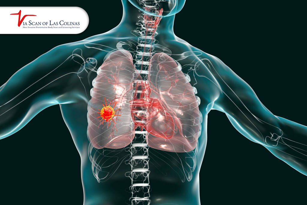
One of the frequent signs of early pancreatic cancer in women is abdominal or back pain. The pancreas is enclosed in the abdomen and is located near the stomach. An enlarged tumor can also pinch on the surrounding nerves and organs, which can be painful. It is generally a sore pain in the stomach and can extend to the back. A significant number of women complain of being in constant pain after consuming food. However, you cannot rule out the possibility that it is being caused by indigestion or stress.
Medical care is needed when it continues to last over two weeks without any improvement. ViaScan CT scan could be used to indicate whether the pancreas is the source of the issue. Our whole body scan provides detailed images that reflect what exactly happens to your body.
Why is unexplained weight loss a sign of pancreatic cancer?
Unexplained weight loss occurs when your body is struggling with a serious issue. It can be a serious sign that you might be struggling with pancreatic cancer. The illness distorts the food processing in the body. However, the pancreas usually secretes enzymes that are very useful in the digestion of food. The presence of a tumor in the pancreas affects the digestion and prevents protein uptake, so even though you can lose weight when you eat normally.
Pancreatic cancer also decreases appetite; you may feel stuffed after every several bites. There are those women who lose ten or more pounds within a couple of months, and they do not know why. Immediately visit a doctor in case you experience unexplained weight loss coupled with abdominal pain or tiredness. The pancreas images are clear in a CT scan at ViaScan. Whole-body scans in the early stages can help in the detection of cancer at the most opportune stage.
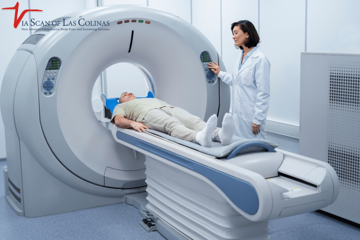
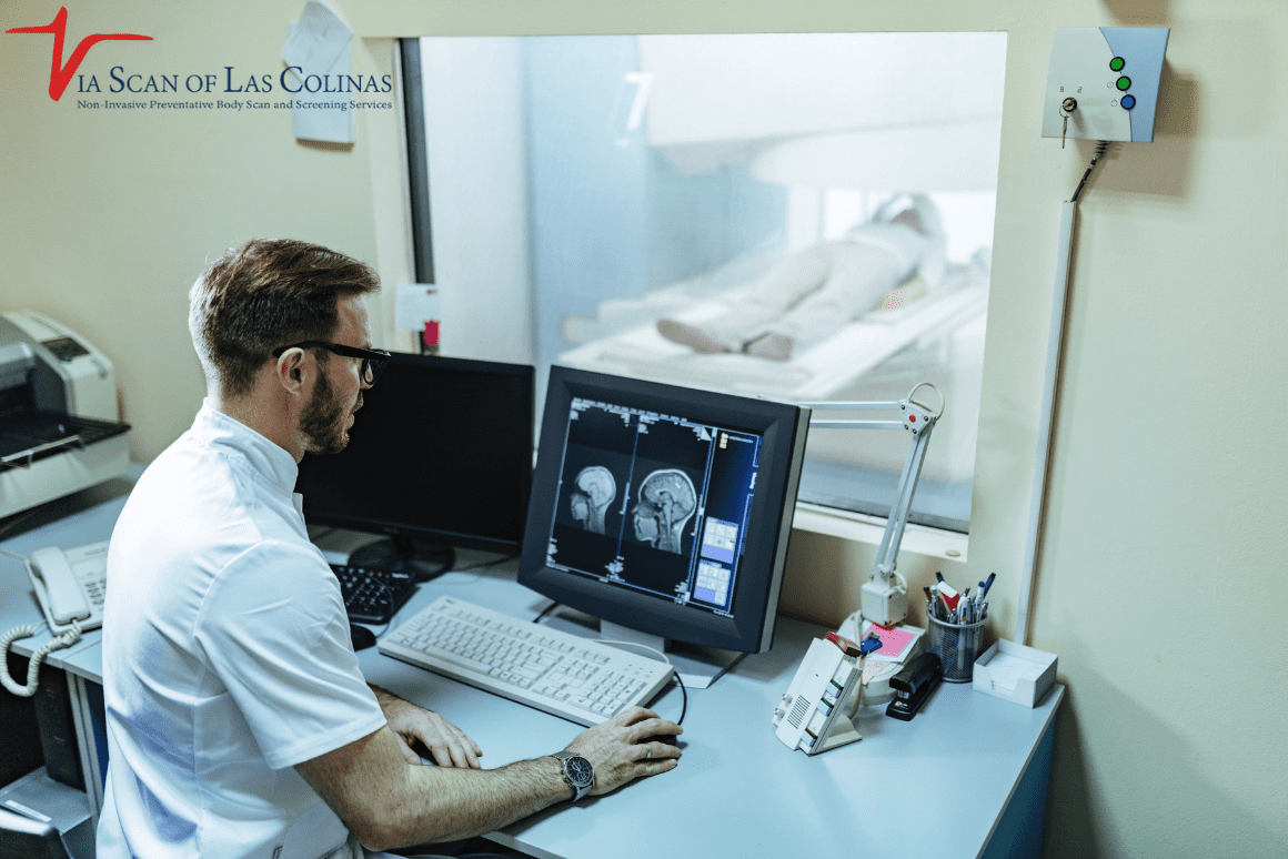
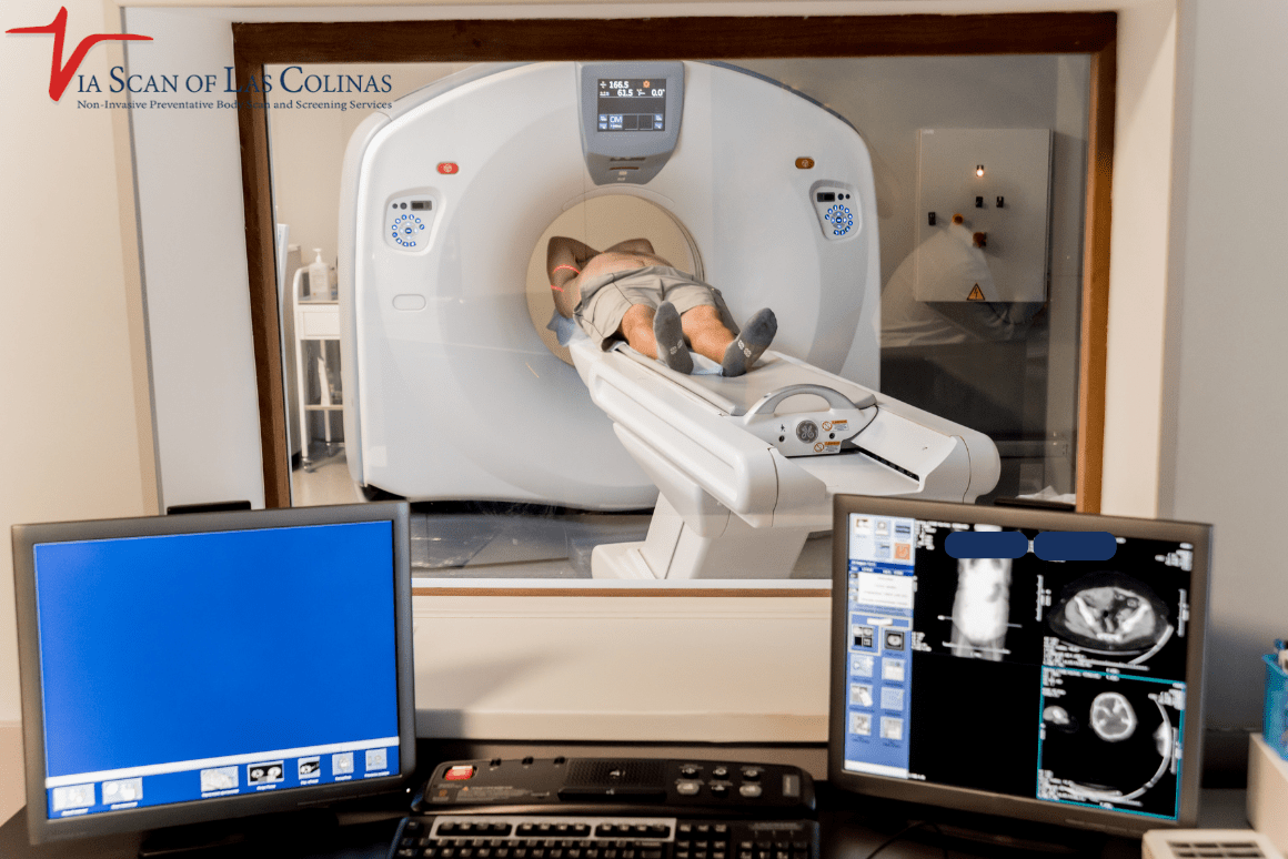
What does the CT scan, or a full body scan, contribute to diagnosing Pancreatic abnormalities?
One of the most effective tools for detecting pancreatic issues at an early onset is a CT scan. A CT scan is an acronym of computed tomography. It is a combination of X-rays and computer programs that create detailed images of your body. The machine gives a picture of your abdomen in numerous different angles. These images expose the pancreas, the surrounding organs, blood vessels, and lymph nodes to ensure that doctors can check for masses, tumors, or irregularities. A complete body scan of the head to pelvis that will show whether the cancer has spread or not. ViaScan is a company that deals with whole-body scanning, providing detailed images of your health.
Choose Our Full Body Scan
Early Detection Saves Lives!
-
- Accurate
- Quick Result
- Affordable

Conclusion
In women, Pancreatic cancer starts causing subtle signs that can be easily disregarded. The symptoms that one should never overlook are abdominal pains, backache, unexplained weight loss, and intestinal issues that persist for several weeks. Women are usually denied or underdiagnosed with their symptoms, and thus, awareness-wise, advocacy is important. Modern imaging, such as a CT scan for pancreatic cancer detection, can be used as a powerful tool in early detection. ViaScan has state-of-the-art equipment and professional scanners to provide whole body scanning. We assist women in finding the right answers in a short period. Your health matters. In case of any concerns, consult with a professional for assistance. Early detection saves lives.